 🔬
Tissue
🔬
Tissue
Pigmentary epi
Eye — Pigmentary epi — Pigmentary epi
Nervous system
Tissue Overview
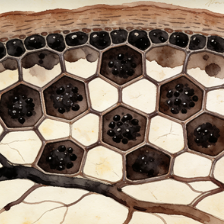
The retinal pigment epithelium (RPE) is a single layer of pigmented cells located between the neural retina and the choroid. It is vital for retinal health: it absorbs stray light, forms part of the blood-retinal barrier, transports nutrients, phagocytoses shed photoreceptor outer segments, and recycles visual pigments (retinoids). RPE dysfunction is implicated in diseases like age-related macular degeneration.
Tissue UMAP Atlas & Gene Explorer
Loading longevity analysis…
Loading UMAP for Pigmentary epi…
Cell Distribution — WebGL
Cells: 0Types: 0Zoom: 1.0x
→ UMAP_1
↑ UMAP_2
Loading...
Cell Types
Loading cell types...
—
Tissues
—
Systems
—
Samples
0
Total Cells
0
Cell Types
Related Tissues
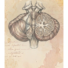
Brain — Cerebellum — Cerebellum
sys01tis09
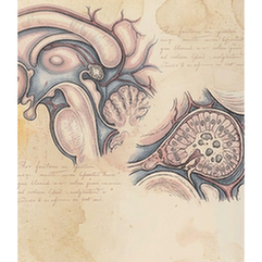
Brain — Pineal gland — Pineal gland
sys01tis14
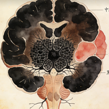
Brain — Substantia nigra — Substantia nigra
sys01tis15
Eye — Pigmentary epi — Pigmentary epi
sys01tis17
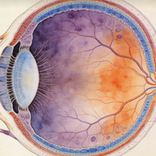
Eye — Retina — Retina
sys01tis18
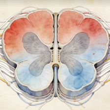
Spinal cord — Spinal cord — Spinal cord
sys01tis20
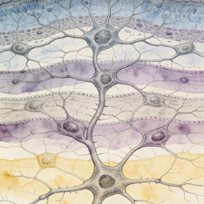
Brain — Neocortex — Neocortex
sys01tis22
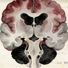
Brain — Brainstem — Midbrain
sys01tis11
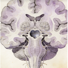
Brain — Brainstem — Pons
sys01tis12
Brain — Olfactory bulb — Olfactory bulb
sys01tis13
Cell Type
Expression
Coords