Tissues
Nervous system
7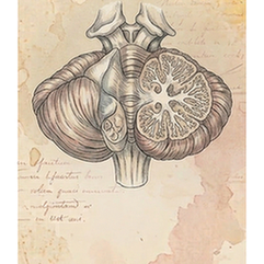
Brain
The cerebellum, or "little brain," is located at the back of the skull below the cerebrum. Despite its size, it contains over half the brain's neurons. Its primary function is coordinating and fine-tuning voluntary movement, ensuring precision, balance, and posture. It also contributes to motor learning, cognitive functions like attention and language, and regulates emotional responses. It does not initiate movement but refines it.
Brain
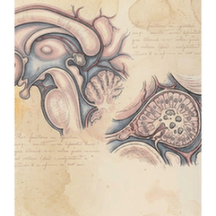
The pineal gland is a small, pine-cone-shaped endocrine gland located deep in the brain, near the center of the two hemispheres. It is best known for producing melatonin, a hormone derived from serotonin that regulates the sleep-wake cycle (circadian rhythm) and seasonal biological rhythms. Its secretion is inhibited by light detected by the retina, making it a crucial neuroendocrine transducer of light-dark information.
Brain
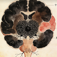
The substantia nigra is a darkly pigmented nucleus located in the midbrain. Its name means "black substance," derived from its high neuromelanin content. It is a critical component of the basal ganglia motor circuit. Its pars compacta region produces dopamine, which is essential for smooth, coordinated movement. The degeneration of these dopaminergic neurons is the primary cause of Parkinson's disease's motor symptoms.
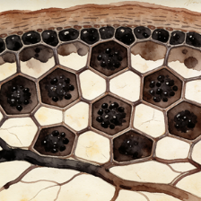
Eye
The retinal pigment epithelium (RPE) is a single layer of pigmented cells located between the neural retina and the choroid. It is vital for retinal health: it absorbs stray light, forms part of the blood-retinal barrier, transports nutrients, phagocytoses shed photoreceptor outer segments, and recycles visual pigments (retinoids). RPE dysfunction is implicated in diseases like age-related macular degeneration.
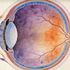
Eye
The retina is the light-sensitive neural tissue lining the back of the eye. It contains photoreceptor cells (rods for dim light, cones for color and detail) that convert light into electrical signals. These signals are processed through a network of bipolar, horizontal, and amacrine cells before being transmitted via retinal ganglion cells, whose axons form the optic nerve, carrying visual information to the brain.
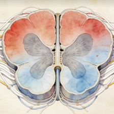
Spinal cord
The spinal cord is a long, cylindrical bundle of nerve tissue that extends from the medulla oblongata down the vertebral column. It serves as the primary conduit for motor information descending from the brain and sensory information ascending to the brain. It also contains central pattern generators for reflexes and coordinated locomotion. It is protected by the vertebrae, meninges, and cerebrospinal fluid.
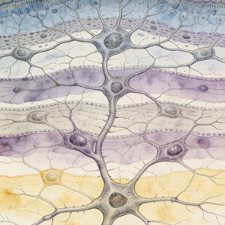
Brain
The neocortex is the evolutionarily newest and largest part of the cerebral cortex, characterized by its six-layered structure. It is responsible for higher brain functions, including sensory perception, generation of motor commands, spatial reasoning, conscious thought, and language. Its extensive, highly folded surface (gyri and sulci) dramatically increases cortical area and cognitive capacity, distinguishing mammals, particularly primates.
Immune system
5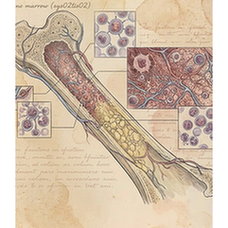
Bone marrow
Bone marrow is the soft, spongy tissue found within the cavities of bones, primarily in the pelvis, sternum, vertebrae, and ribs. It is the primary site of hematopoiesis—the production of all blood cellular components, including red blood cells (erythrocytes), white blood cells (leukocytes), and platelets. It contains hematopoietic stem cells and a supportive stromal microenvironment.
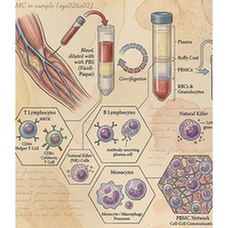
Blood
Peripheral Blood Mononuclear Cells (PBMCs) are a population of blood cells with a single, round nucleus, isolated from peripheral blood. This mixed cell population primarily includes lymphocytes (T cells, B cells, NK cells) and monocytes. They are a critical component of the immune system and are widely used in research and diagnostics for immunophenotyping, functional assays, and genetic studies.
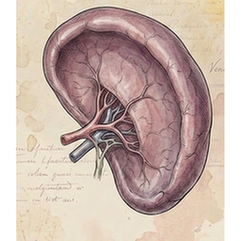
Spleen
The spleen is the largest secondary lymphoid organ, located in the left upper abdomen. It filters blood, removing old or damaged red blood cells and platelets, and captures blood-borne pathogens. Its white pulp contains immune cells (lymphocytes, macrophages) that mount immune responses. The red pulp is responsible for RBC filtration and storage. It is a key site for antibody production and immune surveillance.
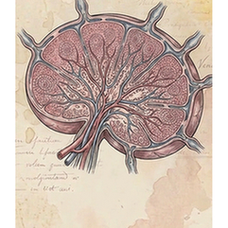
Lymph node
Lymph nodes are small, bean-shaped secondary lymphoid organs distributed along lymphatic vessels throughout the body. They act as filters for lymph fluid, trapping foreign particles and pathogens. They are highly organized structures containing B-cell follicles and T-cell zones, where antigens are presented to lymphocytes, activating adaptive immune responses. Swollen lymph nodes often indicate an active immune response to infection.
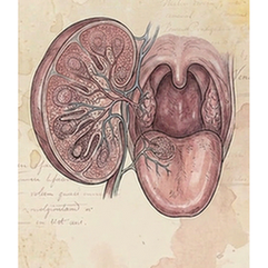
Tonsil
Tonsils are masses of lymphoid tissue located at the back of the throat (oropharynx). They form part of Waldeyer's ring, a first line of immune defense against inhaled or ingested pathogens. Their surface crypts increase area for antigen capture. They contain follicles where B cells are activated, playing a role in mucosal immunity, especially during childhood.
Cardiovascular system
3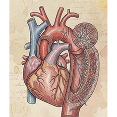
Vessel
The aorta is the largest artery in the body, originating from the left ventricle of the heart. It distributes oxygenated blood to the systemic circulation under high pressure. Its structure includes a thick, elastic wall with three layers (tunica intima, media, adventitia) that allow it to withstand and smooth out the pulsatile flow from the heart. Diseases like aneurysms and atherosclerosis primarily affect the aorta.
Vessel
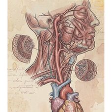
The carotid arteries are major arteries in the neck that supply oxygenated blood to the head and brain. The common carotid divides into the internal carotid (supplying the brain and eyes) and the external carotid (supplying the face and neck). The carotid sinus contains baroreceptors for blood pressure regulation. Carotid atherosclerosis is a major risk factor for ischemic stroke.
Heart
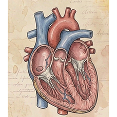
The heart is a muscular organ that functions as the central pump of the circulatory system. It has four chambers: two atria (receiving chambers) and two ventricles (pumping chambers). Through coordinated, rhythmic contractions (the cardiac cycle), it pumps deoxygenated blood to the lungs via the pulmonary circuit and oxygenated blood to the rest of the body via the systemic circuit. Its own blood supply comes from the coronary arteries.
Respiratory system
4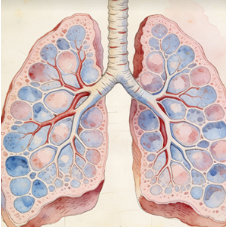
Lung
The lungs are paired, spongy, air-filled organs located in the thoracic cavity. Their primary function is gas exchange: oxygen from inhaled air diffuses into the blood in the alveoli, while carbon dioxide diffuses out to be exhaled. The branching airway tree (trachea to bronchi to bronchioles) conducts air. They also play roles in pH balance, speech, and host defense.
Trachea
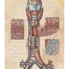
The trachea, or windpipe, is a cartilaginous tube connecting the larynx to the primary bronchi. Its C-shaped hyaline cartilage rings provide rigidity to keep the airway open, while the posterior membranous wall allows flexibility for swallowing. It is lined with ciliated pseudostratified columnar epithelium that traps and moves particles out of the airway. It is the main conduit for air to the lungs.
Bronchus
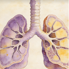
The bronchi are the large airway passages that branch from the trachea into each lung. The right main bronchus is wider and more vertical than the left. They further divide into secondary (lobar) and tertiary (segmental) bronchi, forming the bronchial tree. Their walls contain cartilage plates and smooth muscle. They conduct air to the bronchioles and are involved in conditioning inhaled air.
Diaphragm
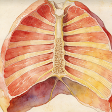
The diaphragm is a large, dome-shaped skeletal muscle that separates the thoracic cavity from the abdominal cavity. It is the primary muscle of inspiration (inhalation). When it contracts, it flattens, increasing thoracic volume and decreasing intra-thoracic pressure, drawing air into the lungs. It is controlled by the phrenic nerves (C3-C5) and also aids in defecation, urination, and childbirth.
Digestive system
9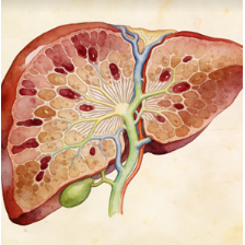
Liver
The liver is the largest internal organ and a metabolic powerhouse located in the upper right abdomen. It performs over 500 vital functions, including detoxification of metabolites, protein synthesis, production of biochemicals for digestion (bile), glycogen storage, regulation of blood glucose, synthesis of plasma proteins (e.g., albumin), and breakdown of old red blood cells. It has a remarkable capacity for regeneration.
Gallbladder
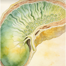
The gallbladder is a small, pear-shaped, muscular sac that stores and concentrates bile produced by the liver. It is located on the inferior surface of the liver. In response to cholecystokinin (CCK) released after a meal, it contracts and releases bile via the cystic and common bile ducts into the duodenum, where bile emulsifies fats to aid in their digestion and absorption.
Stomach
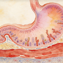
The stomach is a J-shaped muscular sac in the upper abdomen that serves as a temporary reservoir for food. It mechanically churns and chemically digests food via gastric acid (HCl) and enzymes like pepsin. Its lining secretes mucus to protect itself. It regulates the delivery of partially digested food (chyme) into the small intestine. It also produces intrinsic factor, essential for vitamin B12 absorption.
Pancreas
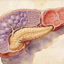
The pancreas is a dual-function gland located behind the stomach. Its exocrine portion (acinar cells) produces digestive enzymes (lipase, amylase, proteases) secreted into the duodenum. Its endocrine portion (islets of Langerhans) secretes hormones directly into the bloodstream, primarily insulin (from beta cells) and glucagon (from alpha cells), which are crucial for blood glucose regulation.
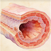
Esophagus
The esophagus is a muscular tube that connects the pharynx to the stomach, passing through the diaphragm. Its primary function is to transport food and liquids from the mouth to the stomach via coordinated, wave-like muscle contractions called peristalsis. The upper and lower esophageal sphincters regulate entry and prevent reflux. Its lining is stratified squamous epithelium for protection against abrasion.
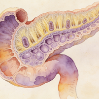
Intestine
The duodenum is the first and shortest segment (about 25 cm) of the small intestine, immediately following the stomach. It is a C-shaped structure where most chemical digestion occurs. It receives chyme from the stomach, bile from the liver/gallbladder, and digestive enzymes from the pancreas. Its Brunner's glands secrete alkaline mucus to neutralize gastric acid, protecting the intestinal lining and optimizing enzyme activity.
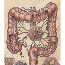
Intestine
The colon, or large intestine, is the final part of the digestive tract. Its main functions are to absorb water and electrolytes from indigestible food matter, compact the remaining material into feces, and store it until defecation. It houses a vast population of commensal bacteria (gut microbiota) that ferment certain substrates and produce vitamins (like K and some B vitamins). It consists of ascending, transverse, descending, and sigmoid sections.
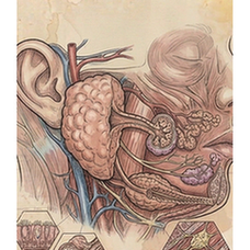
Salivary gland
Salivary glands are exocrine glands that produce saliva, which is secreted into the oral cavity. The three major pairs are the parotid, submandibular, and sublingual glands. Saliva contains water, electrolytes, mucus, antibacterial compounds, and enzymes like amylase (which begins starch digestion). It lubricates food for swallowing, initiates digestion, maintains oral hygiene, and facilitates taste and speech.
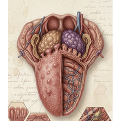
Tongue
The tongue is a muscular organ in the mouth covered with a mucous membrane. Its intrinsic and extrinsic muscles provide precise movement for speech, swallowing, and food manipulation. Its surface is covered with papillae, many of which contain taste buds for gustation (sweet, salty, sour, bitter, umami). It is also crucial for articulating speech sounds and for oral cleansing.
Reproductive system
8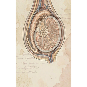
Testis
The testes (singular: testis) are the male primary gonads, housed in the scrotum. They have two primary functions: spermatogenesis, the production of spermatozoa in the seminiferous tubules; and steroidogenesis, the secretion of androgens, primarily testosterone, by Leydig cells. Temperature regulation in the scrotum is essential for normal spermatogenesis. Testosterone drives male secondary sexual characteristics and reproductive function.
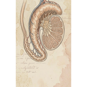
Epididymis
The epididymis is a tightly coiled, comma-shaped tube that sits on the posterior surface of each testis. It is the site of sperm maturation and storage. Immature sperm from the testes enter the head, gain motility and fertilizing capacity as they pass through the body, and are stored in the tail until ejaculation. Its secretions provide an environment for sperm to complete their functional development.
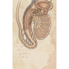
Spermaduct
The spermaduct, more commonly known as the vas deferens or ductus deferens, is a thick-walled, muscular tube that transports mature sperm from the tail of the epididymis to the ejaculatory duct. During ejaculation, its smooth muscle undergoes peristaltic contractions to propel sperm forward. It is a key structure in male reproductive anatomy and is surgically cut or sealed during a vasectomy for male sterilization.
Prostate
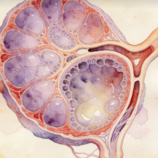
The prostate is a walnut-sized exocrine gland located below the bladder in males. It surrounds the urethra. Its primary function is to secrete a slightly alkaline, milky fluid that constitutes about 30% of semen volume. This fluid contains enzymes (e.g., PSA), citric acid, and zinc, which nourish sperm and enhance their motility and viability. Benign prostatic hyperplasia (BPH) and prostate cancer are common disorders.
Ovary
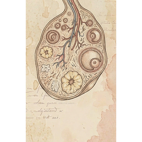
The ovaries are the female primary gonads, located in the pelvic cavity. They have two key functions: gametogenesis, the production and release of oocytes (eggs) through ovulation; and steroidogenesis, the secretion of the sex hormones estrogen and progesterone. These hormones regulate the menstrual cycle, support pregnancy, and maintain female secondary sexual characteristics. The ovaries contain a finite number of follicles at birth.
Fallopian tube
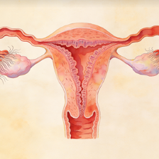
The Fallopian tubes (uterine tubes or oviducts) are paired tubes that extend from near the ovaries to the uterus. They are not directly attached to the ovaries. Their function is to transport the oocyte from the ovary to the uterus. Fertilization by sperm typically occurs in the ampulla region. Their ciliated lining and muscular contractions facilitate the movement of the oocyte or embryo.
Uterus
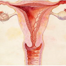
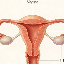
The uterus (womb) is a hollow, muscular, pear-shaped organ located in the female pelvis. It is the site of menstruation, implantation of a fertilized egg, gestation of the fetus, and labor. Its inner lining, the endometrium, cyclically thickens and sheds in response to ovarian hormones. Its thick myometrial wall contracts powerfully during childbirth. The cervix is its lower part, opening into the vagina.
Vagina
The vagina is a fibromuscular, elastic canal that extends from the cervix of the uterus to the external vaginal opening. It serves as the female copulatory organ, the birth canal during parturition, and the passageway for menstrual flow. Its acidic environment, maintained by Lactobacillus species, provides a defense against pathogens. Its walls are lined with stratified squamous epithelium and are highly distensible.
Endocrine system
5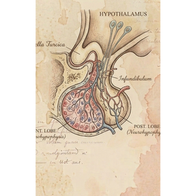
Brain
The pituitary gland, or hypophysis, is a pea-sized endocrine gland located at the base of the brain in the sella turcica. Often called the "master gland," it regulates other endocrine organs. The anterior pituitary (adenohypophysis) secretes tropic hormones (e.g., TSH, ACTH, FSH, LH, GH, Prolactin). The posterior pituitary (neurohypophysis) stores and releases oxytocin and vasopressin (ADH) produced by the hypothalamus.
Adrenal gland
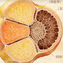
The adrenal glands are small, triangular endocrine glands located atop each kidney. Each gland has two distinct parts: the outer adrenal cortex and the inner adrenal medulla. The cortex produces steroid hormones: mineralocorticoids (aldosterone) for salt/water balance, glucocorticoids (cortisol) for stress and metabolism, and androgens. The medulla secretes catecholamines (epinephrine/adrenaline, norepinephrine) for the "fight-or-flight" response.
Thyroid gland
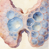
The thyroid gland is a butterfly-shaped endocrine gland located in the anterior neck, below the larynx. It produces thyroid hormones (thyroxine/T4 and triiodothyronine/T3), which are critical for regulating the body's metabolic rate, growth, development, and temperature. It also produces calcitonin, which helps regulate blood calcium levels. Iodine is an essential component of thyroid hormones.
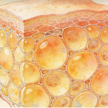
Adipose
Subcutaneous adipose tissue (SAT) is the layer of fat located directly beneath the skin (the hypodermis). It serves as an energy reservoir, provides thermal insulation, and offers mechanical cushioning. Its distribution varies by sex (more in hips/thighs in females, more in abdomen in males). It also acts as an endocrine organ, secreting adipokines like leptin and adiponectin, which influence metabolism and appetite.
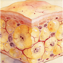
Adipose
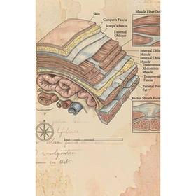
Visceral adipose tissue (VAT) is body fat stored within the abdominal cavity, surrounding internal organs like the liver, pancreas, and intestines. Unlike subcutaneous fat, it is metabolically more active and is strongly associated with increased risk of metabolic diseases, including type 2 diabetes, cardiovascular disease, and insulin resistance. It releases inflammatory cytokines and free fatty acids directly into the portal circulation, affecting liver metabolism.
Urinary system
2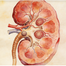
Kidney
The kidneys are paired, bean-shaped organs located retroperitoneally in the upper abdominal area. They are the body's primary filtration system, removing waste products and excess ions from blood to form urine. They critically regulate blood volume, pressure, pH, and electrolyte balance (e.g., sodium, potassium). They also produce hormones: erythropoietin (stimulates RBC production) and renin (regulates blood pressure).
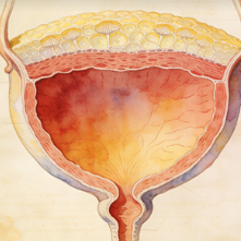
Bladder
The urinary bladder is a hollow, muscular, distensible organ located in the pelvis. Its primary function is to store urine produced by the kidneys before it is excreted from the body via micturition (urination). The detrusor muscle in its wall contracts to expel urine. Its capacity and the sensation of fullness are controlled by complex neural pathways. The internal and external urethral sphincters control release.