Tissues
Nervous system
9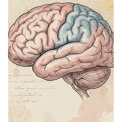
Brain
The frontal lobe is the largest and most anterior lobe of the cerebrum, critical for executive functions. It houses the primary motor cortex, which controls voluntary movements, and the prefrontal cortex, responsible for higher-order cognition such as decision-making, planning, problem-solving, personality expression, and social behavior. Damage to this area can profoundly affect an individual's character and ability to organize thoughts and actions.
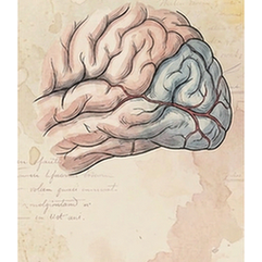
Brain
The occipital lobe is the most posterior region of the cerebrum and is exclusively dedicated to visual processing. It contains the primary visual cortex (V1), which receives raw sensory input from the eyes via the thalamus. Subsequent visual association areas interpret this data, enabling functions like object recognition, color perception, motion detection, and spatial navigation. It is the brain's central hub for making sense of what we see.
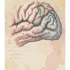
Brain
The parietal lobe, located superior to the occipital lobe and posterior to the frontal lobe, integrates sensory information. Its primary somatosensory cortex processes touch, pressure, temperature, and pain from the body. Association areas are crucial for spatial awareness, proprioception (sense of body position), hand-eye coordination, and complex perceptual functions like reading and arithmetic. It creates a coherent spatial map of the self and the environment.
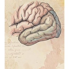
Brain
The temporal lobe lies inferior to the lateral sulcus on each cerebral hemisphere. It is vital for auditory processing via the primary auditory cortex, and for high-level visual processing related to object and face recognition. Its medial region, including the hippocampus, is fundamental for forming and retrieving long-term declarative memories. The left temporal lobe is also central to language comprehension (Wernicke's area).
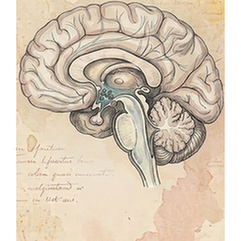
Brain
The hypothalamus is a small but master regulatory region located below the thalamus. It maintains the body's internal balance (homeostasis) by linking the nervous and endocrine systems. It controls autonomic functions like temperature, hunger, thirst, and circadian rhythms. By secreting releasing hormones, it directs the pituitary gland, thus governing growth, metabolism, stress responses, and reproductive functions.
Brain
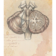
The cerebellum, or "little brain," is located at the back of the skull below the cerebrum. Despite its size, it contains over half the brain's neurons. Its primary function is coordinating and fine-tuning voluntary movement, ensuring precision, balance, and posture. It also contributes to motor learning, cognitive functions like attention and language, and regulates emotional responses. It does not initiate movement but refines it.
Brain
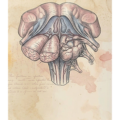
The medulla oblongata is the lowest part of the brainstem, connecting to the spinal cord. It houses vital autonomic control centers essential for survival. These centers regulate involuntary functions such as heart rate (cardiac center), blood pressure (vasomotor center), and the rhythm of breathing (respiratory center). It also serves as a conduit for motor and sensory tracts crossing between the brain and body.
Brain
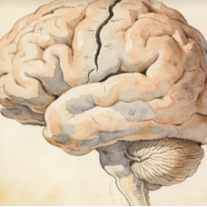
The whole brain refers to the entire encephalon, encompassing all major divisions: cerebrum (cerebral hemispheres), cerebellum, and brainstem (midbrain, pons, medulla). It is the central organ of the nervous system, responsible for integrating sensory information, generating thoughts and emotions, coordinating all voluntary and involuntary bodily functions, and enabling consciousness. It is protected by the skull and meninges.
Eye
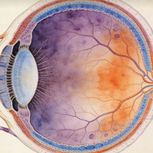
The retina is the light-sensitive neural tissue lining the back of the eye. It contains photoreceptor cells (rods for dim light, cones for color and detail) that convert light into electrical signals. These signals are processed through a network of bipolar, horizontal, and amacrine cells before being transmitted via retinal ganglion cells, whose axons form the optic nerve, carrying visual information to the brain.
Immune system
2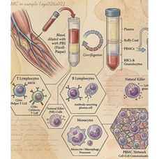
Blood
Peripheral Blood Mononuclear Cells (PBMCs) are a population of blood cells with a single, round nucleus, isolated from peripheral blood. This mixed cell population primarily includes lymphocytes (T cells, B cells, NK cells) and monocytes. They are a critical component of the immune system and are widely used in research and diagnostics for immunophenotyping, functional assays, and genetic studies.
Spleen
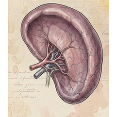
The spleen is the largest secondary lymphoid organ, located in the left upper abdomen. It filters blood, removing old or damaged red blood cells and platelets, and captures blood-borne pathogens. Its white pulp contains immune cells (lymphocytes, macrophages) that mount immune responses. The red pulp is responsible for RBC filtration and storage. It is a key site for antibody production and immune surveillance.
Digestive system
3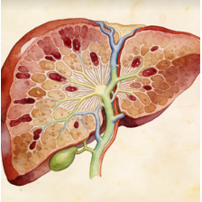
Liver
The liver is the largest internal organ and a metabolic powerhouse located in the upper right abdomen. It performs over 500 vital functions, including detoxification of metabolites, protein synthesis, production of biochemicals for digestion (bile), glycogen storage, regulation of blood glucose, synthesis of plasma proteins (e.g., albumin), and breakdown of old red blood cells. It has a remarkable capacity for regeneration.
Intestine
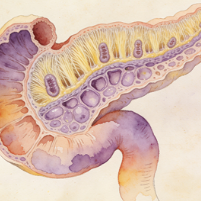
The duodenum is the first and shortest segment (about 25 cm) of the small intestine, immediately following the stomach. It is a C-shaped structure where most chemical digestion occurs. It receives chyme from the stomach, bile from the liver/gallbladder, and digestive enzymes from the pancreas. Its Brunner's glands secrete alkaline mucus to neutralize gastric acid, protecting the intestinal lining and optimizing enzyme activity.