Cell Types
Neural cells
5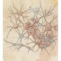
Neural progenitor cells
Neural progenitor cells are mitotically active precursors that generate neurons and glial cells during nervous system development and, in restricted niches such as the subventricular zone and the hippocampal dentate gyrus, in the adult brain. They balance self-renewal and differentiation through tightly regulated signaling pathways. Their decline with age is associated with reduced neurogenesis.
Enteric glial cells
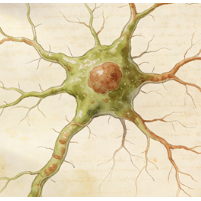
Enteric glial cells (EGCs) are the resident glia of the enteric nervous system (ENS), the "gut brain." They resemble CNS astrocytes and are integral to gastrointestinal homeostasis. EGCs support enteric neurons, regulate intestinal barrier integrity, modulate immune responses within the gut wall, and influence motility. Their dysfunction is implicated in inflammatory bowel diseases (IBD), infections, and functional GI disorders, highlighting their role beyond mere structural support.
Enteric neuronal progenitor cells
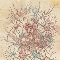
Enteric neuronal progenitor cells (ENPCs) are stem/precursor cells that give rise to the neurons and glia of the enteric nervous system (ENS). Primarily active during embryonic development, they migrate, proliferate, and differentiate to colonize the entire gut. Some progenitor-like cells may persist in adults, offering potential for ENS plasticity and repair. Defects in their migration cause Hirschsprung's disease, a congenital absence of ENS neurons in the distal colon.
Melanocytes
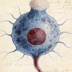
Melanocytes are neural crest-derived cells located in the basal layer of the epidermis and in hair follicles. Their primary function is the production of melanin, a pigment that provides color to skin and hair and protects underlying cells from UV radiation damage. Melanin is synthesized in organelles called melanosomes, which are then transferred to neighboring keratinocytes via dendritic processes.
Neuronal cells
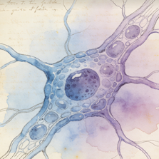
Neuronal cells (Neural cells) in species SP115. Single-cell UMAP atlas.
Immune cells
5
B cells
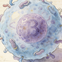
B cells are lymphocytes responsible for the humoral arm of the adaptive immune response. They develop in the bone marrow and, upon maturation, circulate in blood and lymph. When activated by antigen (often with T cell help), they proliferate and differentiate into antibody-secreting plasma cells or long-lived memory B cells. Their antibodies neutralize pathogens, opsonize them for phagocytosis, and activate the complement system.
Dendritic cells
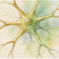
Dendritic cells (DCs) are professional antigen-presenting cells (APCs) that act as sentinels linking innate and adaptive immunity. Immature DCs in tissues capture antigens, then mature and migrate to lymph nodes. There, they present processed antigens on MHC molecules to naive T cells, providing the critical "signal 1" and costimulatory "signal 2" required for T cell activation and differentiation, thereby initiating antigen-specific immune responses.
Macrophages
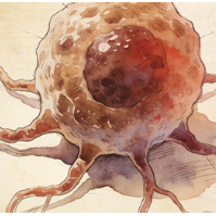
Macrophages are large, phagocytic cells of the innate immune system present in all tissues (where they have specific names like Kupffer cells, microglia). They derive from blood monocytes or local progenitors. They engulf and destroy pathogens and dead cells, secrete cytokines and chemokines to regulate inflammation, present antigens, and are key players in tissue repair, remodeling, and homeostasis. They exhibit remarkable functional plasticity (M1/M2 spectra).
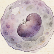
Monocytes
Monocytes are circulating white blood cells (agranulocytes) that serve as precursors for macrophages and dendritic cells. Produced in the bone marrow, they patrol the bloodstream for several days before migrating into tissues in response to inflammatory signals. In tissues, they differentiate into macrophages or dendritic cells. In blood, they can phagocytose pathogens and present antigens, acting as a bridge between innate and adaptive immunity.
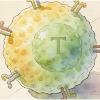
T cells
T cells (T lymphocytes) are central players in cell-mediated adaptive immunity. They develop in the thymus and express T cell receptors (TCRs) that recognize peptide antigens presented by MHC molecules. Major subsets include: Helper T cells (CD4+, which secrete cytokines to help B cells and macrophages), Cytotoxic T cells (CD8+, which kill infected/cancerous cells), and Regulatory T cells (Tregs, which suppress immune responses to prevent autoimmunity).
Epithelial cells
11
Acinar cells
Acinar cells are the exocrine secretory cells of glands like the salivary glands and pancreas. In the pancreas, they are organized into acini and synthesize, store, and secrete digestive pro-enzymes (zymogens) into a ductal network. They have a highly developed rough ER and abundant secretory (zymogen) granules. When stimulated (e.g., by cholecystokinin), they release their contents into the pancreatic duct, which empties into the duodenum.
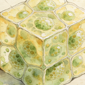
Alveolar type 2 cells
Alveolar type 2 (AT2) cells are cuboidal epithelial cells found in the alveolar corners. They have three critical functions: (1) They synthesize and secrete pulmonary surfactant, a phospholipid-protein mixture that reduces surface tension and prevents alveolar collapse. (2) They serve as progenitor cells for both AT1 and AT2 cells, repairing the alveolar epithelium after injury. (3) They contribute to innate immune defense in the alveoli.
Ascending loop of Henle cells
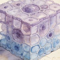
Cells of the thick ascending limb (TAL) of the loop of Henle in the kidney nephron. They are impermeable to water but actively reabsorb sodium, potassium, and chloride via the Na-K-2Cl cotransporter (NKCC2) on their apical membrane. This transport is critical for generating the hypertonic medullary interstitium necessary for water reabsorption in the collecting duct. They also contribute to magnesium and calcium reabsorption.
Cholangiocytes
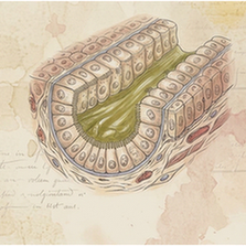
Cholangiocytes are the epithelial cells that line the intrahepatic and extrahepatic bile ducts. They modify the composition of bile (through secretion and absorption of water, ions, and bicarbonate), form a protective barrier, and respond to hormonal signals. They express receptors and channels (e.g., CFTR) critical for bile flow. Their proliferation and dysfunction are central to cholangiopathies like primary sclerosing cholangitis (PSC).
Ciliated cells
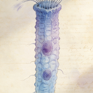
Ciliated cells are epithelial cells characterized by the presence of motile cilia (hair-like projections) on their apical surface. They are found in the respiratory tract, oviducts, and ventricles of the brain (ependymal cells). Coordinated, rhythmic beating of cilia moves fluid or mucus over the epithelial surface. In the airways, this "mucociliary escalator" traps and propels inhaled particles and pathogens out of the lungs, a key defense mechanism.
Enterocytes
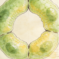
Enterocytes are the principal absorptive columnar epithelial cells lining the small intestine villi. Their apical surface is covered with microvilli, forming a "brush border" that dramatically increases surface area for nutrient absorption. They digest and absorb carbohydrates, peptides, amino acids, and lipids. They also secrete water and ions and form a crucial barrier between the gut lumen and the internal environment.
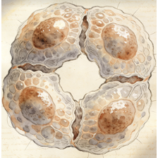
Hepatocytes
Hepatocytes are the principal parenchymal cells of the liver, making up about 80% of its mass. They are metabolic powerhouses with functions including: protein synthesis (albumin, clotting factors), bile production, detoxification and metabolism of drugs/toxins, glycogen storage and glucose homeostasis, lipid metabolism, and cholesterol synthesis. They are arranged in plates radiating from central veins, with polarized faces facing sinusoids and bile canaliculi.
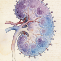
Podocytes
Podocytes are highly specialized epithelial cells in the Bowman's capsule of the kidney glomerulus. They wrap around capillaries with primary and secondary foot processes. The slits between these foot processes, covered by the slit diaphragm, form the final filtration barrier preventing proteins from entering the urine. Podocyte injury or loss leads to proteinuria and glomerular diseases like focal segmental glomerulosclerosis (FSGS).
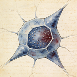
Proximal tubule cells
These cells line the proximal convoluted tubule (PCT) of the nephron, the first and longest segment. They are responsible for the bulk reabsorption of filtered nutrients (glucose, amino acids), ions (sodium, chloride, bicarbonate), and water (~65%). Their apical surface has a dense brush border of microvilli to increase surface area. They also actively secrete organic acids and bases into the filtrate.
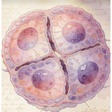
Squamous cells
Squamous cells are thin, flat, scale-like epithelial cells. Simple squamous epithelium (e.g., endothelium, alveolar lining) facilitates diffusion and filtration. Stratified squamous epithelium (e.g., skin epidermis, oral mucosa, esophagus) provides protection against abrasion and pathogens. The outermost cells may be keratinized (skin) or non-keratinized (esophagus). Their shape minimizes friction and allows for easy sloughing and replacement.
Urothelial cells
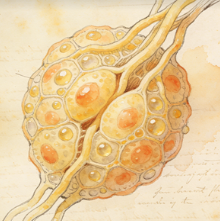
Urothelial cells (transitional epithelial cells) line the urinary tract from renal pelvis to urethra. They form a distensible, impermeable barrier. The superficial umbrella cells have a unique apical plaque of uroplakin proteins and can change shape (from cuboidal to squamous) as the bladder fills and stretches, preventing urine from leaking into underlying tissues. This epithelium also protects against toxic substances in urine.
Stromal cells
3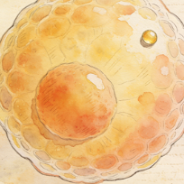
Adipocytes
Adipocytes (fat cells) are the primary cells of adipose tissue, specialized in storing energy as triglycerides within a large, single lipid droplet (white adipocytes) or in dissipating energy as heat via uncoupling protein 1 (UCP1) in numerous smaller droplets (brown/beige adipocytes). White adipocytes also secrete hormones (leptin, adiponectin) and cytokines, making adipose tissue an important endocrine organ involved in metabolism and inflammation.
Fibroblasts
Fibroblasts are the most common cells of connective tissue. They synthesize and secrete the extracellular matrix (ECM) components, including collagen, elastin, and fibronectin, providing structural and biochemical support to surrounding cells. They are key players in wound healing, where they proliferate, migrate to the injury site, and differentiate into contractile myofibroblasts to close wounds. They also communicate with immune and epithelial cells.
Stromal cells
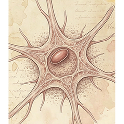
Stromal cells in human. 101,901 cells across 16 tissues.
Muscle cells
2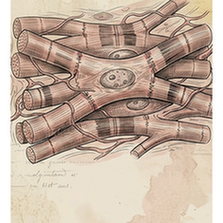
Cardiomyocytes
Cardiomyocytes are the striated muscle cells that constitute the cardiac muscle of the heart. They are responsible for generating the contractile force that pumps blood. They are connected end-to-end by intercalated discs containing gap junctions for electrical coupling and desmosomes for mechanical attachment. Unlike skeletal muscle, they are mononucleated or binucleated and have intrinsic automaticity (pacemaker cells).
Smooth muscle cells
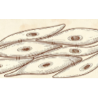
Smooth muscle cells (SMCs) are involuntary, non-striated muscle cells found in the walls of hollow organs (e.g., blood vessels, gut, bladder, uterus). They are spindle-shaped with a single central nucleus. They contract slowly and can maintain tension for long periods (tonus). Contraction is regulated by the autonomic nervous system, hormones, and local factors (e.g., nitric oxide, endothelin).
Other
4
Mesothelial cells
Mesothelial cells form a monolayer (the mesothelium) that lines the body's serous cavities (pleural, pericardial, peritoneal) and covers the outer surface of internal organs. They produce a lubricating serous fluid that allows organs to move smoothly. They also provide a protective barrier, participate in immune surveillance and inflammation, and can undergo transition to a fibroblast-like phenotype (mesothelial-to-mesenchymal transition) in pathology.
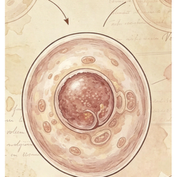
Proliferating cells
Proliferating cells (Other) in species SP115. Single-cell UMAP atlas.
Unknown cell type
Unknown cell type (Other) in species SP115. Single-cell UMAP atlas.
Unknown/Uncertain Cell Type
Unknown/Uncertain Cell Type (Other) in species SP115. Single-cell UMAP atlas.