Tissues
Nervous system
12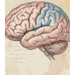
Brain
The frontal lobe is the largest and most anterior lobe of the cerebrum, critical for executive functions. It houses the primary motor cortex, which controls voluntary movements, and the prefrontal cortex, responsible for higher-order cognition such as decision-making, planning, problem-solving, personality expression, and social behavior. Damage to this area can profoundly affect an individual's character and ability to organize thoughts and actions.
Brain
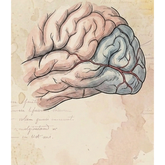
The occipital lobe is the most posterior region of the cerebrum and is exclusively dedicated to visual processing. It contains the primary visual cortex (V1), which receives raw sensory input from the eyes via the thalamus. Subsequent visual association areas interpret this data, enabling functions like object recognition, color perception, motion detection, and spatial navigation. It is the brain's central hub for making sense of what we see.
Brain
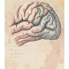
The parietal lobe, located superior to the occipital lobe and posterior to the frontal lobe, integrates sensory information. Its primary somatosensory cortex processes touch, pressure, temperature, and pain from the body. Association areas are crucial for spatial awareness, proprioception (sense of body position), hand-eye coordination, and complex perceptual functions like reading and arithmetic. It creates a coherent spatial map of the self and the environment.
Brain
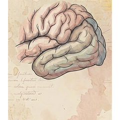
The temporal lobe lies inferior to the lateral sulcus on each cerebral hemisphere. It is vital for auditory processing via the primary auditory cortex, and for high-level visual processing related to object and face recognition. Its medial region, including the hippocampus, is fundamental for forming and retrieving long-term declarative memories. The left temporal lobe is also central to language comprehension (Wernicke's area).
Brain
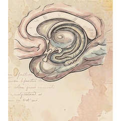
The hippocampus is a curved, seahorse-shaped structure located within the medial temporal lobe. It is the core component of the brain's limbic system and is indispensable for memory formation. Specifically, it consolidates short-term memories into long-term storage and is crucial for spatial memory and navigation. Its vulnerability makes it a primary focus in studies of Alzheimer's disease and amnesia.
Brain
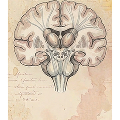
The thalamus is a paired, egg-shaped structure deep within the brain, acting as the brain's major relay station. Nearly all sensory information (except smell) passes through it, where it is filtered and directed to the appropriate cortical areas for conscious perception. It also regulates consciousness, sleep, alertness, and relays motor signals from the cerebellum and basal ganglia to the motor cortex.
Brain
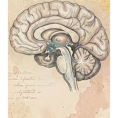
The hypothalamus is a small but master regulatory region located below the thalamus. It maintains the body's internal balance (homeostasis) by linking the nervous and endocrine systems. It controls autonomic functions like temperature, hunger, thirst, and circadian rhythms. By secreting releasing hormones, it directs the pituitary gland, thus governing growth, metabolism, stress responses, and reproductive functions.
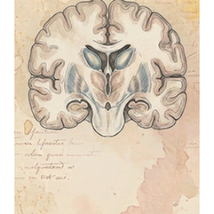
Brain
The striatum is a major input nucleus of the basal ganglia, a group of subcortical structures. Composed of the caudate nucleus and putamen, it receives extensive input from the cerebral cortex. It is critical for facilitating voluntary, purposeful movement and for procedural learning, habit formation, and reward-motivated behavior. Its dysfunction is central to movement disorders like Parkinson's and Huntington's disease.
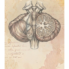
Brain
The cerebellum, or "little brain," is located at the back of the skull below the cerebrum. Despite its size, it contains over half the brain's neurons. Its primary function is coordinating and fine-tuning voluntary movement, ensuring precision, balance, and posture. It also contributes to motor learning, cognitive functions like attention and language, and regulates emotional responses. It does not initiate movement but refines it.
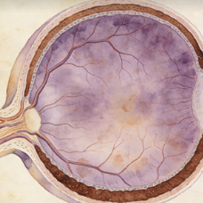
Eye
This complex refers to the functional unit of the posterior eye, comprising the neural retina and the underlying retinal pigment epithelium (RPE). The neural retina detects and processes light, while the RPE provides critical metabolic support, waste removal, and light absorption. Their intimate structural and functional relationship is essential for normal vision; diseases often involve both layers.
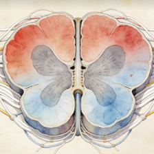
Spinal cord
The spinal cord is a long, cylindrical bundle of nerve tissue that extends from the medulla oblongata down the vertebral column. It serves as the primary conduit for motor information descending from the brain and sensory information ascending to the brain. It also contains central pattern generators for reflexes and coordinated locomotion. It is protected by the vertebrae, meninges, and cerebrospinal fluid.
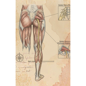
Sciatic nerve
The sciatic nerve is the largest and longest peripheral nerve in the human body. It originates from the lumbosacral plexus (L4-S3) in the lower back and runs down the posterior thigh, branching into the tibial and common fibular nerves near the knee. It provides motor innervation to the muscles of the posterior thigh, leg, and foot, and carries sensory information from these regions.
Immune system
5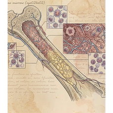
Bone marrow
Bone marrow is the soft, spongy tissue found within the cavities of bones, primarily in the pelvis, sternum, vertebrae, and ribs. It is the primary site of hematopoiesis—the production of all blood cellular components, including red blood cells (erythrocytes), white blood cells (leukocytes), and platelets. It contains hematopoietic stem cells and a supportive stromal microenvironment.
Blood
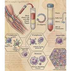
Peripheral Blood Mononuclear Cells (PBMCs) are a population of blood cells with a single, round nucleus, isolated from peripheral blood. This mixed cell population primarily includes lymphocytes (T cells, B cells, NK cells) and monocytes. They are a critical component of the immune system and are widely used in research and diagnostics for immunophenotyping, functional assays, and genetic studies.
Spleen
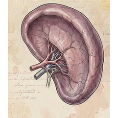
The spleen is the largest secondary lymphoid organ, located in the left upper abdomen. It filters blood, removing old or damaged red blood cells and platelets, and captures blood-borne pathogens. Its white pulp contains immune cells (lymphocytes, macrophages) that mount immune responses. The red pulp is responsible for RBC filtration and storage. It is a key site for antibody production and immune surveillance.
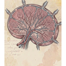
Lymph node
Lymph nodes are small, bean-shaped secondary lymphoid organs distributed along lymphatic vessels throughout the body. They act as filters for lymph fluid, trapping foreign particles and pathogens. They are highly organized structures containing B-cell follicles and T-cell zones, where antigens are presented to lymphocytes, activating adaptive immune responses. Swollen lymph nodes often indicate an active immune response to infection.
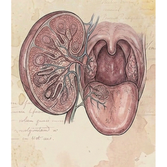
Tonsil
Tonsils are masses of lymphoid tissue located at the back of the throat (oropharynx). They form part of Waldeyer's ring, a first line of immune defense against inhaled or ingested pathogens. Their surface crypts increase area for antigen capture. They contain follicles where B cells are activated, playing a role in mucosal immunity, especially during childhood.
Cardiovascular system
2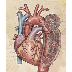
Vessel
The aorta is the largest artery in the body, originating from the left ventricle of the heart. It distributes oxygenated blood to the systemic circulation under high pressure. Its structure includes a thick, elastic wall with three layers (tunica intima, media, adventitia) that allow it to withstand and smooth out the pulsatile flow from the heart. Diseases like aneurysms and atherosclerosis primarily affect the aorta.
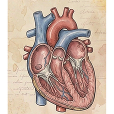
Heart
The heart is a muscular organ that functions as the central pump of the circulatory system. It has four chambers: two atria (receiving chambers) and two ventricles (pumping chambers). Through coordinated, rhythmic contractions (the cardiac cycle), it pumps deoxygenated blood to the lungs via the pulmonary circuit and oxygenated blood to the rest of the body via the systemic circuit. Its own blood supply comes from the coronary arteries.
Digestive system
9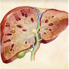
Liver
The liver is the largest internal organ and a metabolic powerhouse located in the upper right abdomen. It performs over 500 vital functions, including detoxification of metabolites, protein synthesis, production of biochemicals for digestion (bile), glycogen storage, regulation of blood glucose, synthesis of plasma proteins (e.g., albumin), and breakdown of old red blood cells. It has a remarkable capacity for regeneration.
Gallbladder
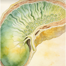
The gallbladder is a small, pear-shaped, muscular sac that stores and concentrates bile produced by the liver. It is located on the inferior surface of the liver. In response to cholecystokinin (CCK) released after a meal, it contracts and releases bile via the cystic and common bile ducts into the duodenum, where bile emulsifies fats to aid in their digestion and absorption.
Stomach
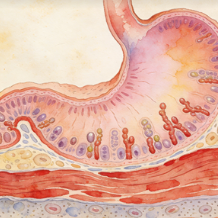
The stomach is a J-shaped muscular sac in the upper abdomen that serves as a temporary reservoir for food. It mechanically churns and chemically digests food via gastric acid (HCl) and enzymes like pepsin. Its lining secretes mucus to protect itself. It regulates the delivery of partially digested food (chyme) into the small intestine. It also produces intrinsic factor, essential for vitamin B12 absorption.
Pancreas
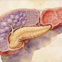
The pancreas is a dual-function gland located behind the stomach. Its exocrine portion (acinar cells) produces digestive enzymes (lipase, amylase, proteases) secreted into the duodenum. Its endocrine portion (islets of Langerhans) secretes hormones directly into the bloodstream, primarily insulin (from beta cells) and glucagon (from alpha cells), which are crucial for blood glucose regulation.
Esophagus
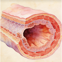
The esophagus is a muscular tube that connects the pharynx to the stomach, passing through the diaphragm. Its primary function is to transport food and liquids from the mouth to the stomach via coordinated, wave-like muscle contractions called peristalsis. The upper and lower esophageal sphincters regulate entry and prevent reflux. Its lining is stratified squamous epithelium for protection against abrasion.
Intestine
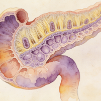
The duodenum is the first and shortest segment (about 25 cm) of the small intestine, immediately following the stomach. It is a C-shaped structure where most chemical digestion occurs. It receives chyme from the stomach, bile from the liver/gallbladder, and digestive enzymes from the pancreas. Its Brunner's glands secrete alkaline mucus to neutralize gastric acid, protecting the intestinal lining and optimizing enzyme activity.
Intestine
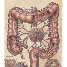
The colon, or large intestine, is the final part of the digestive tract. Its main functions are to absorb water and electrolytes from indigestible food matter, compact the remaining material into feces, and store it until defecation. It houses a vast population of commensal bacteria (gut microbiota) that ferment certain substrates and produce vitamins (like K and some B vitamins). It consists of ascending, transverse, descending, and sigmoid sections.
Salivary gland
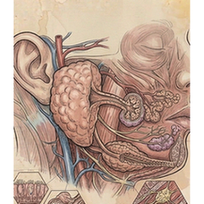
Salivary glands are exocrine glands that produce saliva, which is secreted into the oral cavity. The three major pairs are the parotid, submandibular, and sublingual glands. Saliva contains water, electrolytes, mucus, antibacterial compounds, and enzymes like amylase (which begins starch digestion). It lubricates food for swallowing, initiates digestion, maintains oral hygiene, and facilitates taste and speech.
Tongue
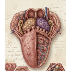
The tongue is a muscular organ in the mouth covered with a mucous membrane. Its intrinsic and extrinsic muscles provide precise movement for speech, swallowing, and food manipulation. Its surface is covered with papillae, many of which contain taste buds for gustation (sweet, salty, sour, bitter, umami). It is also crucial for articulating speech sounds and for oral cleansing.
Endocrine system
4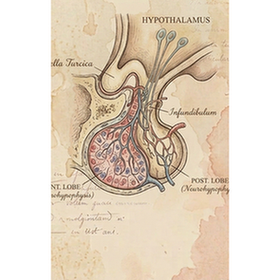
Brain
The pituitary gland, or hypophysis, is a pea-sized endocrine gland located at the base of the brain in the sella turcica. Often called the "master gland," it regulates other endocrine organs. The anterior pituitary (adenohypophysis) secretes tropic hormones (e.g., TSH, ACTH, FSH, LH, GH, Prolactin). The posterior pituitary (neurohypophysis) stores and releases oxytocin and vasopressin (ADH) produced by the hypothalamus.
Adrenal gland
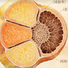
The adrenal glands are small, triangular endocrine glands located atop each kidney. Each gland has two distinct parts: the outer adrenal cortex and the inner adrenal medulla. The cortex produces steroid hormones: mineralocorticoids (aldosterone) for salt/water balance, glucocorticoids (cortisol) for stress and metabolism, and androgens. The medulla secretes catecholamines (epinephrine/adrenaline, norepinephrine) for the "fight-or-flight" response.
Thyroid gland
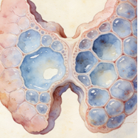
The thyroid gland is a butterfly-shaped endocrine gland located in the anterior neck, below the larynx. It produces thyroid hormones (thyroxine/T4 and triiodothyronine/T3), which are critical for regulating the body's metabolic rate, growth, development, and temperature. It also produces calcitonin, which helps regulate blood calcium levels. Iodine is an essential component of thyroid hormones.
Adipose
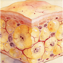
Visceral adipose tissue (VAT) is body fat stored within the abdominal cavity, surrounding internal organs like the liver, pancreas, and intestines. Unlike subcutaneous fat, it is metabolically more active and is strongly associated with increased risk of metabolic diseases, including type 2 diabetes, cardiovascular disease, and insulin resistance. It releases inflammatory cytokines and free fatty acids directly into the portal circulation, affecting liver metabolism.
Motor system
3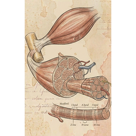
Muscle
Skeletal muscle is a type of striated muscle tissue attached to bones by tendons. It is under voluntary control via the somatic nervous system and is responsible for all locomotion, posture, and voluntary movements. Each muscle fiber is multinucleated and contains highly organized myofibrils with sarcomeres, the contractile units. It also functions as a major metabolic organ and an endocrine tissue, secreting myokines.
Cartilage
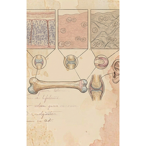
Cartilage is a resilient and flexible connective tissue found in various parts of the body. Its extracellular matrix is rich in collagen fibers and proteoglycans, providing tensile strength and compression resistance. Major types include hyaline cartilage (joint surfaces, ribs, respiratory tract), elastic cartilage (ear, epiglottis), and fibrocartilage (intervertebral discs, menisci). It is avascular and aneural, receiving nutrients by diffusion.
Abdominal wall
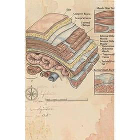
The abdominal wall is a multi-layered structure that encloses the abdominal cavity. Its layers, from superficial to deep, typically include skin, subcutaneous tissue, muscles (external oblique, internal oblique, transversus abdominis, rectus abdominis), fascia, and parietal peritoneum. It provides core support and protection for abdominal viscera, enables trunk movement, maintains posture, and assists in functions like respiration, defecation, and childbirth.
Urinary system
2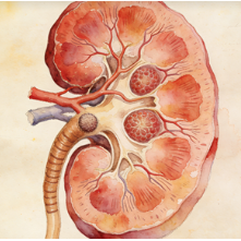
Kidney
The kidneys are paired, bean-shaped organs located retroperitoneally in the upper abdominal area. They are the body's primary filtration system, removing waste products and excess ions from blood to form urine. They critically regulate blood volume, pressure, pH, and electrolyte balance (e.g., sodium, potassium). They also produce hormones: erythropoietin (stimulates RBC production) and renin (regulates blood pressure).
Bladder
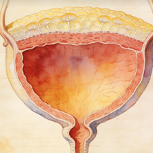
The urinary bladder is a hollow, muscular, distensible organ located in the pelvis. Its primary function is to store urine produced by the kidneys before it is excreted from the body via micturition (urination). The detrusor muscle in its wall contracts to expel urine. Its capacity and the sensation of fullness are controlled by complex neural pathways. The internal and external urethral sphincters control release.