Cell Types
Neural cells
13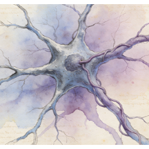
Astrocytes
Astrocytes are star-shaped glial cells, the most abundant in the central nervous system (CNS). They are vital for homeostasis: they form part of the blood-brain barrier, regulate blood flow, control extracellular ion/neurotransmitter balance (e.g., glutamate uptake), and provide metabolic support to neurons. Through the "tripartite synapse," they modulate synaptic strength and plasticity. They also respond to injury via reactive astrogliosis, playing dual roles in scar formation and neuroprotection.
Bipolar cells
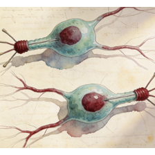
Bipolar cells are retinal neurons that relay visual information directly from photoreceptors (rods and cones) to ganglion cells. They have a characteristic morphology with one dendrite connecting to photoreceptors and one axon synapsing in the inner plexiform layer. They are functionally divided into ON and OFF types, responding to light increments and decrements, respectively. This segregation initiates the parallel pathways essential for contrast perception and visual processing.
Cone cells
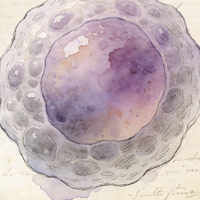
Cone cells are photoreceptors specialized for bright-light (photopic) vision, high visual acuity, and color perception. They are concentrated in the fovea centralis. Humans possess three types, each containing a photopigment sensitive to short (blue), medium (green), or long (red) wavelengths, enabling trichromatic color vision. Cones are less light-sensitive than rods but provide fine spatial detail and are critical for tasks like reading, driving, and recognizing faces and colors.
Enteric glial cells
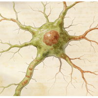
Enteric glial cells (EGCs) are the resident glia of the enteric nervous system (ENS), the "gut brain." They resemble CNS astrocytes and are integral to gastrointestinal homeostasis. EGCs support enteric neurons, regulate intestinal barrier integrity, modulate immune responses within the gut wall, and influence motility. Their dysfunction is implicated in inflammatory bowel diseases (IBD), infections, and functional GI disorders, highlighting their role beyond mere structural support.
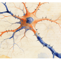
Excitatory neurons
Excitatory neurons are nerve cells that, when activated, increase the likelihood of their target neuron firing an action potential. They primarily release the neurotransmitter glutamate at their synapses. They form the primary driving force in neural circuits throughout the brain (e.g., cortical pyramidal neurons) and are fundamental for information propagation, synaptic plasticity (like LTP), and processes such as learning, memory, and cognition.
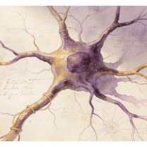
Ganglion cells
Ganglion cells are the output neurons of the retina. Their axons bundle together to form the optic nerve, transmitting processed visual information to the brain (thalamus, hypothalamus, midbrain). Different subtypes (e.g., M/P cells in primates) encode specific visual features like motion, fine detail, or color. Intrinsically photosensitive retinal ganglion cells (ipRGCs) contain melanopsin and regulate non-image-forming functions like circadian rhythms and pupillary reflex.
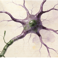
Inhibitory neurons
Inhibitory neurons reduce the probability of their postsynaptic target neuron firing an action potential. They primarily release the neurotransmitters GABA (in the brain) or glycine (in the spinal cord/brainstem). They are crucial for balancing neural circuit activity, preventing runaway excitation, generating rhythmic outputs (e.g., in pacemaker circuits), and shaping the temporal and spatial precision of neural computations. Types include interneurons like basket cells and Purkinje cells.
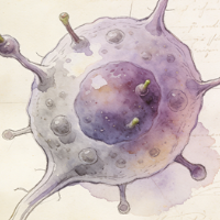
Oligodendrocyte progenitor cells
Oligodendrocyte progenitor cells (OPCs), also called NG2-glia, are a population of glial cells found throughout the CNS. They are proliferative and maintain the capacity to differentiate into mature, myelinating oligodendrocytes. OPCs are critical for developmental myelination and remain in the adult CNS, where they contribute to remyelination after demyelinating injuries (e.g., in Multiple Sclerosis) and may have additional roles in monitoring neural circuits.
Oligodendrocytes
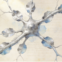
Oligodendrocytes are the myelinating glial cells of the central nervous system (CNS). Each oligodendrocyte extends multiple processes to wrap and insulate segments of several different axons with concentric layers of myelin membrane. This myelin sheath greatly increases the speed of action potential propagation (saltatory conduction). They also provide metabolic support to the axons they enwrap. Loss of oligodendrocytes or myelin disrupts nerve signaling.
Rod cells
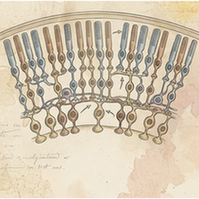
Rod cells are photoreceptors specialized for low-light (scotopic) vision. They are extremely light-sensitive, containing the pigment rhodopsin, and are responsible for night vision and peripheral vision. Rods do not mediate color perception, providing monochromatic (black-and-white) vision. Their signals converge heavily onto bipolar cells, maximizing sensitivity at the expense of fine detail. They are more numerous than cones and are absent from the central fovea.
Schwann cells
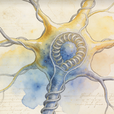
Schwann cells are the principal glial cells of the peripheral nervous system (PNS). Myelinating Schwann cells wrap around single axons to form the myelin sheath, enabling fast saltatory conduction. Non-myelinating Schwann cells ensheath multiple small-diameter axons. Schwann cells are crucial for supporting axon health and guiding regeneration after PNS injury, forming regeneration tubes (Bands of Büngner) that direct axonal regrowth.
Microglia
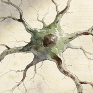
Microglia are the resident macrophages and primary immune cells of the central nervous system (CNS). In their resting state, they constantly survey the parenchyma. Upon detecting injury, infection, or pathological signals, they activate, becoming phagocytic to clear debris, dead cells, and pathogens. They also play essential roles in synaptic pruning during development, modulating neuroinflammation, and can contribute to both repair and neurodegeneration in diseases.
Melanocytes
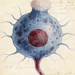
Melanocytes are neural crest-derived cells located in the basal layer of the epidermis and in hair follicles. Their primary function is the production of melanin, a pigment that provides color to skin and hair and protects underlying cells from UV radiation damage. Melanin is synthesized in organelles called melanosomes, which are then transferred to neighboring keratinocytes via dendritic processes.
Immune cells
17
B cells
B cells are lymphocytes responsible for the humoral arm of the adaptive immune response. They develop in the bone marrow and, upon maturation, circulate in blood and lymph. When activated by antigen (often with T cell help), they proliferate and differentiate into antibody-secreting plasma cells or long-lived memory B cells. Their antibodies neutralize pathogens, opsonize them for phagocytosis, and activate the complement system.
Cycling T cells
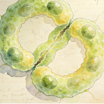
Cycling T cells are T lymphocytes undergoing active cell division. This is a hallmark of T cell activation following antigen recognition via the TCR and costimulation. Massive clonal expansion occurs, transforming a few naive antigen-specific T cells into thousands of effector T cells to combat infection. It is a fundamental step in mounting an effective adaptive immune response and generating long-lived memory T cells.
Dendritic cells
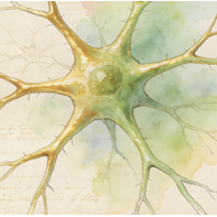
Dendritic cells (DCs) are professional antigen-presenting cells (APCs) that act as sentinels linking innate and adaptive immunity. Immature DCs in tissues capture antigens, then mature and migrate to lymph nodes. There, they present processed antigens on MHC molecules to naive T cells, providing the critical "signal 1" and costimulatory "signal 2" required for T cell activation and differentiation, thereby initiating antigen-specific immune responses.
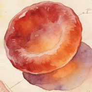
Erythroid cells
Erythroid cells represent the lineage of hematopoietic cells committed to becoming erythrocytes (red blood cells). This includes progenitors (BFU-E, CFU-E) and morphologically identifiable precursors: proerythroblasts, basophilic, polychromatophilic, and orthochromatic erythroblasts, which undergo hemoglobin synthesis, nuclear condensation, and finally enucleation to form reticulocytes and then mature RBCs. Their primary function is oxygen transport via hemoglobin.
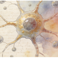
Hematopoietic stem and progenitor cells
Hematopoietic stem and progenitor cells (HSPCs) reside in the bone marrow and are responsible for lifelong blood cell production (hematopoiesis). Hematopoietic stem cells (HSCs) have long-term self-renewal and multipotent differentiation capacity. Progenitor cells (like CMPs, CLPs) are more restricted in potential but highly proliferative. Together, they give rise to all lineages of blood and immune cells.
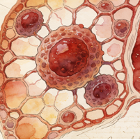
Kupffer cells
Kupffer cells are specialized tissue-resident macrophages located within the sinusoids of the liver. They are the largest population of fixed macrophages in the body. They phagocytose pathogens, toxins, cellular debris, and aged red blood cells from the portal blood flow. They play crucial roles in liver immunity, iron recycling, lipid metabolism, and maintaining overall hepatic homeostasis. Their activation can contribute to liver inflammation and fibrosis.
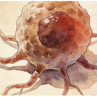
Macrophages
Macrophages are large, phagocytic cells of the innate immune system present in all tissues (where they have specific names like Kupffer cells, microglia). They derive from blood monocytes or local progenitors. They engulf and destroy pathogens and dead cells, secrete cytokines and chemokines to regulate inflammation, present antigens, and are key players in tissue repair, remodeling, and homeostasis. They exhibit remarkable functional plasticity (M1/M2 spectra).
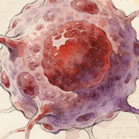
Megakaryocyte erythrocyte progenitors
Megakaryocyte erythrocyte progenitors (MEPs) are bipotent hematopoietic progenitor cells derived from the Common Myeloid Progenitor (CMP). MEPs are committed to producing two critical blood cell types: megakaryocytes (which fragment into platelets for clotting) and erythrocytes (red blood cells for oxygen transport). The MEP stage represents the final branch point before lineage-specific commitment to platelet or red cell production.
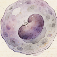
Monocytes
Monocytes are circulating white blood cells (agranulocytes) that serve as precursors for macrophages and dendritic cells. Produced in the bone marrow, they patrol the bloodstream for several days before migrating into tissues in response to inflammatory signals. In tissues, they differentiate into macrophages or dendritic cells. In blood, they can phagocytose pathogens and present antigens, acting as a bridge between innate and adaptive immunity.
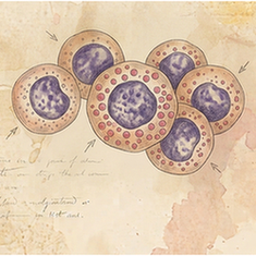
Natural killer cells
Natural Killer (NK) cells are cytotoxic lymphocytes of the innate immune system. They provide rapid responses to virus-infected cells and tumor cells without prior sensitization. They use a balance of activating and inhibitory receptors to detect "missing self" (loss of MHC I) or "induced self" (stress ligands). They kill targets by releasing perforin and granzymes (cytotoxic granules) and through death receptor ligands like FasL.
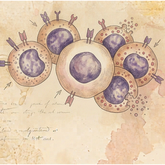
Natural killer T cells
Natural Killer T (NKT) cells are a unique subset of T lymphocytes that bridge innate and adaptive immunity. They express an invariant T cell receptor (TCR) that recognizes lipid antigens presented by the non-classical MHC molecule CD1d. Upon activation, they rapidly produce large quantities of cytokines (e.g., IFN-γ, IL-4), influencing the activity of many other immune cells and shaping immune responses to infection, cancer, and autoimmunity.
Neutrophils
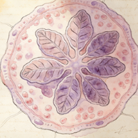
Neutrophils are the most abundant white blood cell and the first responders to sites of bacterial or fungal infection. They are highly motile phagocytic cells that engulf and destroy pathogens using antimicrobial granules (containing myeloperoxidase, defensins) and reactive oxygen species. They form neutrophil extracellular traps (NETs) to ensnare microbes. Their short lifespan and potent effector functions make them crucial for acute inflammation.
Plasma B cells
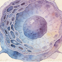
Plasma B cells (or plasma cells) are the terminal, fully differentiated effector state of activated B lymphocytes. They are antibody factories, specializing in the massive secretion of antibodies of a single specificity. They possess extensive endoplasmic reticulum and a prominent Golgi apparatus to support high-rate protein synthesis. Most are short-lived, but some become long-lived plasma cells that reside in the bone marrow, providing sustained antibody levels.
Platelets
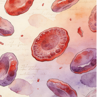
Platelets (thrombocytes) are small, anucleate cell fragments derived from megakaryocytes in the bone marrow. They circulate in blood and are essential for hemostasis: upon vascular injury, they adhere, activate, aggregate to form a plug, and provide a surface for coagulation factors. They also secrete growth factors and cytokines, playing roles in inflammation, immunity, and tissue repair.
T cells
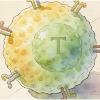
T cells (T lymphocytes) are central players in cell-mediated adaptive immunity. They develop in the thymus and express T cell receptors (TCRs) that recognize peptide antigens presented by MHC molecules. Major subsets include: Helper T cells (CD4+, which secrete cytokines to help B cells and macrophages), Cytotoxic T cells (CD8+, which kill infected/cancerous cells), and Regulatory T cells (Tregs, which suppress immune responses to prevent autoimmunity).
Goblet cells
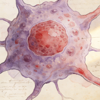
Goblet cells are specialized unicellular mucus-secreting glands found within simple columnar epithelia of the respiratory and intestinal tracts. They have a characteristic goblet shape, with a basal nucleus and an apical region packed with mucin granules. They secrete mucins, which hydrate and form the protective mucus layer that traps particles, lubricates surfaces, and protects the epithelium from pathogens and damage.
Pancreas islet cells
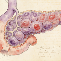
Pancreatic islet (islets of Langerhans) cells are the endocrine cells of the pancreas, organized into clusters scattered throughout the exocrine tissue. Major types include: Alpha cells (secrete glucagon, raises blood sugar), Beta cells (secrete insulin, lowers blood sugar), Delta cells (secrete somatostatin, inhibits other islet cells), and PP cells (secrete pancreatic polypeptide). They precisely regulate glucose homeostasis.
Epithelial cells
25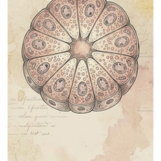
Acinar cells
Acinar cells are the exocrine secretory cells of glands like the salivary glands and pancreas. In the pancreas, they are organized into acini and synthesize, store, and secrete digestive pro-enzymes (zymogens) into a ductal network. They have a highly developed rough ER and abundant secretory (zymogen) granules. When stimulated (e.g., by cholecystokinin), they release their contents into the pancreatic duct, which empties into the duodenum.
Alveolar type 1 cells
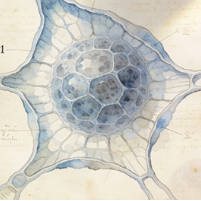
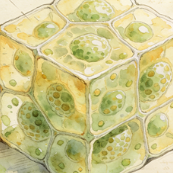
Alveolar type 1 (AT1) cells are thin, squamous epithelial cells that cover approximately 95% of the gas exchange surface in the lung alveoli. Their extremely flattened morphology (extending over large areas) is ideal for the passive diffusion of oxygen and carbon dioxide between the airspace and the underlying pulmonary capillaries. They are terminally differentiated and crucial for maintaining the delicate blood-air barrier.
Alveolar type 2 cells
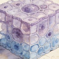
Alveolar type 2 (AT2) cells are cuboidal epithelial cells found in the alveolar corners. They have three critical functions: (1) They synthesize and secrete pulmonary surfactant, a phospholipid-protein mixture that reduces surface tension and prevents alveolar collapse. (2) They serve as progenitor cells for both AT1 and AT2 cells, repairing the alveolar epithelium after injury. (3) They contribute to innate immune defense in the alveoli.
Ascending loop of Henle cells
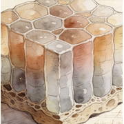
Cells of the thick ascending limb (TAL) of the loop of Henle in the kidney nephron. They are impermeable to water but actively reabsorb sodium, potassium, and chloride via the Na-K-2Cl cotransporter (NKCC2) on their apical membrane. This transport is critical for generating the hypertonic medullary interstitium necessary for water reabsorption in the collecting duct. They also contribute to magnesium and calcium reabsorption.
Basal cells
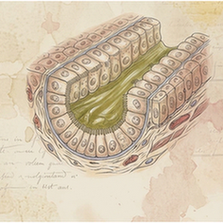
Basal cells are a layer of mitotically active epithelial progenitor/stem cells found attached to the basement membrane in stratified and pseudostratified epithelia (e.g., skin, airways, prostate). They divide to give rise to cells that differentiate and move upward to replenish the superficial layers. In the airway, they are major stem cells capable of regenerating both ciliated and secretory cells after injury.
Cholangiocytes
Cholangiocytes are the epithelial cells that line the intrahepatic and extrahepatic bile ducts. They modify the composition of bile (through secretion and absorption of water, ions, and bicarbonate), form a protective barrier, and respond to hormonal signals. They express receptors and channels (e.g., CFTR) critical for bile flow. Their proliferation and dysfunction are central to cholangiopathies like primary sclerosing cholangitis (PSC).
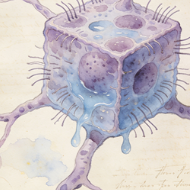
Choroid plexus
The choroid plexus is a specialized structure in the brain's ventricles composed of a network of capillaries surrounded by a monolayer of modified epithelial cells (choroid plexus epithelial cells). Its primary function is to produce cerebrospinal fluid (CSF) by filtering blood plasma and actively secreting ions and water. It also forms the blood-CSF barrier and secretes growth factors and signaling molecules into the CSF.
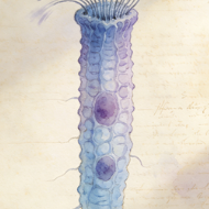
Ciliated cells
Ciliated cells are epithelial cells characterized by the presence of motile cilia (hair-like projections) on their apical surface. They are found in the respiratory tract, oviducts, and ventricles of the brain (ependymal cells). Coordinated, rhythmic beating of cilia moves fluid or mucus over the epithelial surface. In the airways, this "mucociliary escalator" traps and propels inhaled particles and pathogens out of the lungs, a key defense mechanism.
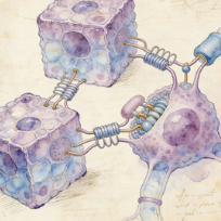
Connecting tubule cells
Connecting tubule (CNT) cells are specialized cells in the nephron that link the distal convoluted tubule to the collecting duct. They play a significant role in fine-tuning sodium, potassium, and acid-base balance. They are sensitive to aldosterone and reabsorb sodium while secreting potassium and protons. They express the calcium channel TRPV5 and are important for the final regulation of calcium reabsorption.
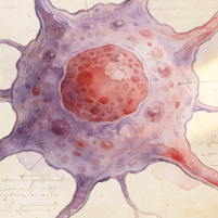
Corticotropes
Corticotropes are endocrine cells in the anterior pituitary gland (adenohypophysis) that produce and secrete adrenocorticotropic hormone (ACTH). ACTH is derived from the precursor pro-opiomelanocortin (POMC). In response to corticotropin-releasing hormone (CRH) from the hypothalamus, corticotropes release ACTH into the bloodstream, which then stimulates the adrenal cortex to produce glucocorticoids (like cortisol), key stress hormones.
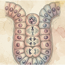
Cycling epithelial cells
A broad categorization for any epithelial cell (from various tissues) that is captured in an active phase of the cell cycle. This label is used in genomic datasets to distinguish proliferating cells from quiescent or terminally differentiated ones. It highlights the dynamic nature of epithelia, where constant renewal and repair are driven by these cycling populations.
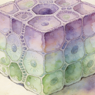
Distal convoluted tubule cells
Cells of the distal convoluted tubule (DCT) in the nephron. They are responsible for fine-tuning electrolyte balance. They actively reabsorb sodium and chloride (via the Na-Cl cotransporter, NCC) and are a major site for parathyroid hormone (PTH)-regulated calcium reabsorption. Their activity is crucial for maintaining blood pressure, potassium, calcium, and magnesium homeostasis.
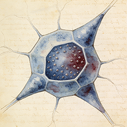
Endocrine cells
Endocrine cells (Epithelial cells) in species SP301. Single-cell UMAP atlas.
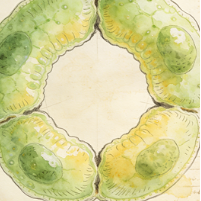
Enterocytes
Enterocytes are the principal absorptive columnar epithelial cells lining the small intestine villi. Their apical surface is covered with microvilli, forming a "brush border" that dramatically increases surface area for nutrient absorption. They digest and absorb carbohydrates, peptides, amino acids, and lipids. They also secrete water and ions and form a crucial barrier between the gut lumen and the internal environment.
Ependymal cells
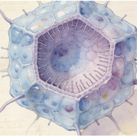
Ependymal cells are ciliated epithelial-like glial cells that line the ventricles of the brain and the central canal of the spinal cord. Their coordinated ciliary beating helps circulate cerebrospinal fluid (CSF). They contribute to the blood-CSF barrier, secrete factors into the CSF, and in certain niches (like the lateral ventricles), some can act as neural stem cells (radial glia-like cells).
Glandular cells
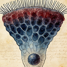
Glandular cells are specialized epithelial cells that constitute the secretory units of exocrine and endocrine glands. They synthesize and release specific products. Exocrine glandular cells (e.g., in salivary, sweat glands) secrete via ducts. Endocrine glandular cells (e.g., in thyroid, pituitary) secrete hormones directly into the bloodstream. Their morphology is adapted for secretion, with abundant rough ER and secretory granules.
Hepatocytes
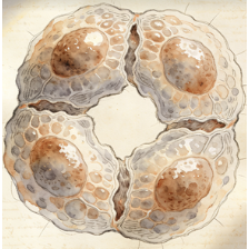
Hepatocytes are the principal parenchymal cells of the liver, making up about 80% of its mass. They are metabolic powerhouses with functions including: protein synthesis (albumin, clotting factors), bile production, detoxification and metabolism of drugs/toxins, glycogen storage and glucose homeostasis, lipid metabolism, and cholesterol synthesis. They are arranged in plates radiating from central veins, with polarized faces facing sinusoids and bile canaliculi.
Keratinocytes
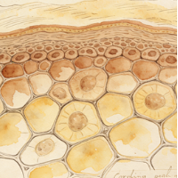
Keratinocytes are the predominant cell type in the epidermis (the outermost skin layer). They originate from basal layer stem cells and undergo a process of terminal differentiation as they move outward, producing keratin intermediate filaments. In the stratum corneum, they become anucleate corneocytes, forming a tough, waterproof barrier that protects against mechanical injury, water loss, and microbial invasion.
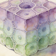
Luminal epithelial cells
These are the epithelial cells that directly face the lumen (hollow cavity) of a duct or gland. In tissues like the mammary gland or prostate, they form the inner secretory layer and are often hormonally responsive. They are typically more differentiated than basal cells and carry out the tissue-specific secretory functions (e.g., producing milk proteins or prostatic fluid).
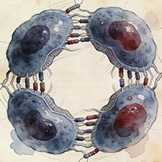
Pancreas ductal cells
Pancreatic ductal cells form the lining of the ductal network that transports digestive enzymes from the acini and bicarbonate-rich fluid from the centroacinar cells to the duodenum. They modify the pancreatic juice by secreting bicarbonate ions (HCO3-) to neutralize gastric acid. They express CFTR, and mutations here cause cystic fibrosis-related pancreatic disease. They can also act as progenitor cells in certain contexts.
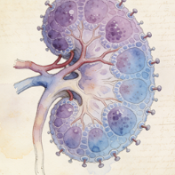
Podocytes
Podocytes are highly specialized epithelial cells in the Bowman's capsule of the kidney glomerulus. They wrap around capillaries with primary and secondary foot processes. The slits between these foot processes, covered by the slit diaphragm, form the final filtration barrier preventing proteins from entering the urine. Podocyte injury or loss leads to proteinuria and glomerular diseases like focal segmental glomerulosclerosis (FSGS).
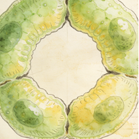
Principal cells
In the kidney's collecting duct, principal cells are the most abundant cell type. They are responsible for the final regulation of water, sodium, and potassium balance. They express aquaporin-2 channels on their apical membrane (inserted in response to vasopressin/ADH) for water reabsorption. They also reabsorb sodium via ENaC channels and secrete potassium, processes regulated by aldosterone.
Proximal tubule cells
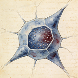
These cells line the proximal convoluted tubule (PCT) of the nephron, the first and longest segment. They are responsible for the bulk reabsorption of filtered nutrients (glucose, amino acids), ions (sodium, chloride, bicarbonate), and water (~65%). Their apical surface has a dense brush border of microvilli to increase surface area. They also actively secrete organic acids and bases into the filtrate.
Serous cells
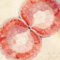
Serous cells are protein-secreting epithelial cells found in exocrine glands like the salivary glands (parotid) and pancreatic acini. Their secretory granules contain enzymes (e.g., amylase in saliva, digestive enzymes in pancreas). The granules often stain darkly with H&E. They typically have a round nucleus and basophilic cytoplasm due to abundant rough ER for protein synthesis.
Urothelial cells
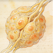
Urothelial cells (transitional epithelial cells) line the urinary tract from renal pelvis to urethra. They form a distensible, impermeable barrier. The superficial umbrella cells have a unique apical plaque of uroplakin proteins and can change shape (from cuboidal to squamous) as the bladder fills and stretches, preventing urine from leaking into underlying tissues. This epithelium also protects against toxic substances in urine.
Stromal cells
6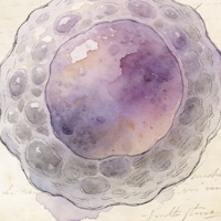
Adipocyte progenitor cells
Adipocyte progenitor cells (APCs), also known as preadipocytes, are mesenchymal stem cells committed to the adipocyte lineage. They reside in adipose tissue depots and have the capacity to proliferate and differentiate into mature lipid-filled adipocytes. They play key roles in adipose tissue expansion, both in normal growth and in obesity. They also secrete various factors (adipokines) and contribute to tissue remodeling.
Adipocytes
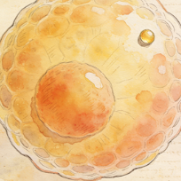
Adipocytes (fat cells) are the primary cells of adipose tissue, specialized in storing energy as triglycerides within a large, single lipid droplet (white adipocytes) or in dissipating energy as heat via uncoupling protein 1 (UCP1) in numerous smaller droplets (brown/beige adipocytes). White adipocytes also secrete hormones (leptin, adiponectin) and cytokines, making adipose tissue an important endocrine organ involved in metabolism and inflammation.
Fibroblasts
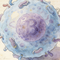
Fibroblasts are the most common cells of connective tissue. They synthesize and secrete the extracellular matrix (ECM) components, including collagen, elastin, and fibronectin, providing structural and biochemical support to surrounding cells. They are key players in wound healing, where they proliferate, migrate to the injury site, and differentiate into contractile myofibroblasts to close wounds. They also communicate with immune and epithelial cells.
Other stromal cells
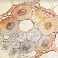
Stromal cells are a heterogeneous group of connective tissue cells that provide structural support and create the microenvironment ("stroma") for parenchymal cells within an organ. They include fibroblasts, adipocytes, pericytes, and various types of mesenchymal stem/stromal cells. They produce ECM, secrete growth factors and cytokines, and play critical roles in tissue architecture, homeostasis, repair, and disease progression (e.g., in cancer).
Pericytes
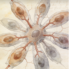
Pericytes are contractile mesenchymal cells that wrap around the endothelial cells of capillaries and venules. They are embedded in the basement membrane and are crucial for vascular stability, regulating blood flow, promoting endothelial survival, and participating in the formation and maintenance of the blood-brain barrier. They also have stem cell properties and can differentiate into other mesenchymal cells during tissue repair.
Stellate cells
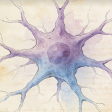
In the liver, hepatic stellate cells (HSCs) reside in the space of Disse. In their quiescent state, they store vitamin A. Upon liver injury, they activate, proliferate, and transform into myofibroblast-like cells that produce large amounts of extracellular matrix, driving liver fibrosis and cirrhosis. Pancreatic stellate cells play a similar fibrogenic role in pancreatitis and pancreatic cancer.
Muscle cells
5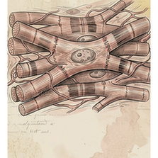
Cardiomyocytes
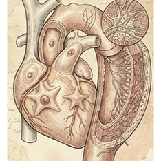
Cardiomyocytes are the striated muscle cells that constitute the cardiac muscle of the heart. They are responsible for generating the contractile force that pumps blood. They are connected end-to-end by intercalated discs containing gap junctions for electrical coupling and desmosomes for mechanical attachment. Unlike skeletal muscle, they are mononucleated or binucleated and have intrinsic automaticity (pacemaker cells).
Satellite cells
Satellite cells are the resident stem cells of skeletal muscle, located between the basal lamina and the sarcolemma of muscle fibers. They are normally quiescent but activate, proliferate, and differentiate into myoblasts in response to injury or exercise. They fuse with existing fibers or with each other to form new myofibers, enabling muscle growth and repair.
Skeletal myonuclei
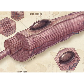
Skeletal myonuclei are the multiple nuclei contained within a single, multinucleated skeletal muscle fiber (myofiber). They are post-mitotic and are responsible for gene expression to maintain the massive cytoplasmic volume of the myofiber. They are evenly distributed, but in some muscle types, they may be clustered near the neuromuscular junction or myotendinous junction.
Smooth muscle cells
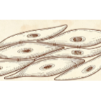
Smooth muscle cells (SMCs) are involuntary, non-striated muscle cells found in the walls of hollow organs (e.g., blood vessels, gut, bladder, uterus). They are spindle-shaped with a single central nucleus. They contract slowly and can maintain tension for long periods (tonus). Contraction is regulated by the autonomic nervous system, hormones, and local factors (e.g., nitric oxide, endothelin).
Type II myonuclei
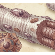
In skeletal muscle, myonuclei within fast-twitch (Type II) muscle fibers. These fibers are glycolytic, generate more force but fatigue quickly. Type II myonuclei (subtypes IIa, IIx/d) govern the expression of genes for glycolytic enzymes, fast myosin heavy chains, and other components defining the fast, anaerobic metabolic phenotype of the fiber.
Germ cells
3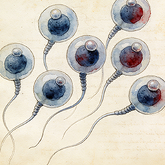
Spermatids
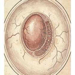
Spermatocytes are developing male germ cells that undergo meiosis. Primary spermatocytes (diploid, 4N DNA) result from spermatogonial mitosis. They undergo Meiosis I to form two secondary spermatocytes (haploid, 2N DNA). Secondary spermatocytes rapidly undergo Meiosis II to produce four spermatids (haploid, 1N DNA). This process occurs within the seminiferous tubules, supported by Sertoli cells.
Spermatocytes
Spermatocytes are developing male germ cells that undergo meiosis. Primary spermatocytes (diploid, 4N DNA) result from spermatogonial mitosis. They undergo Meiosis I to form two secondary spermatocytes (haploid, 2N DNA). Secondary spermatocytes rapidly undergo Meiosis II to produce four spermatids (haploid, 1N DNA). This process occurs within the seminiferous tubules, supported by Sertoli cells.
Spermatogonia
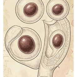
Spermatogonia are the undifferentiated germ stem cells located on the basement membrane of the seminiferous tubules. They undergo mitotic divisions to self-renew and produce a population of cells committed to differentiation (primary spermatocytes). Type A spermatogonia are the stem cell reservoir, while Type B spermatogonia are committed progenitors. They are the foundation of lifelong sperm production.
Other
3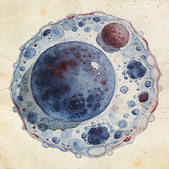
Promyelocytes
Promyelocytes (Other) in species SP301. Single-cell UMAP atlas.
Intercalated cells
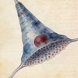
Intercalated cells are specialized cells found in the collecting ducts of the kidney nephron. They are critical for regulating systemic acid-base balance. There are two main types: Type A (alpha) cells secrete protons (H+) via H+-ATPase pumps and reabsorb bicarbonate, correcting acidosis. Type B (beta) cells secrete bicarbonate and reabsorb protons, correcting alkalosis. They also play a role in potassium balance.
Mesothelial cells
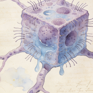
Mesothelial cells form a monolayer (the mesothelium) that lines the body's serous cavities (pleural, pericardial, peritoneal) and covers the outer surface of internal organs. They produce a lubricating serous fluid that allows organs to move smoothly. They also provide a protective barrier, participate in immune surveillance and inflammation, and can undergo transition to a fibroblast-like phenotype (mesothelial-to-mesenchymal transition) in pathology.