Tissues
Nervous system
3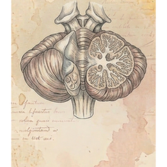
Brain
The cerebellum, or "little brain," is located at the back of the skull below the cerebrum. Despite its size, it contains over half the brain's neurons. Its primary function is coordinating and fine-tuning voluntary movement, ensuring precision, balance, and posture. It also contributes to motor learning, cognitive functions like attention and language, and regulates emotional responses. It does not initiate movement but refines it.
Brain
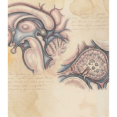
The pineal gland is a small, pine-cone-shaped endocrine gland located deep in the brain, near the center of the two hemispheres. It is best known for producing melatonin, a hormone derived from serotonin that regulates the sleep-wake cycle (circadian rhythm) and seasonal biological rhythms. Its secretion is inhibited by light detected by the retina, making it a crucial neuroendocrine transducer of light-dark information.
Brain
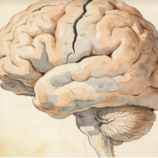
The whole brain refers to the entire encephalon, encompassing all major divisions: cerebrum (cerebral hemispheres), cerebellum, and brainstem (midbrain, pons, medulla). It is the central organ of the nervous system, responsible for integrating sensory information, generating thoughts and emotions, coordinating all voluntary and involuntary bodily functions, and enabling consciousness. It is protected by the skull and meninges.
Immune system
2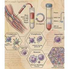
Blood
Peripheral Blood Mononuclear Cells (PBMCs) are a population of blood cells with a single, round nucleus, isolated from peripheral blood. This mixed cell population primarily includes lymphocytes (T cells, B cells, NK cells) and monocytes. They are a critical component of the immune system and are widely used in research and diagnostics for immunophenotyping, functional assays, and genetic studies.
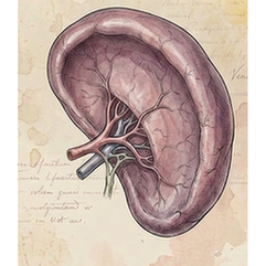
Spleen
The spleen is the largest secondary lymphoid organ, located in the left upper abdomen. It filters blood, removing old or damaged red blood cells and platelets, and captures blood-borne pathogens. Its white pulp contains immune cells (lymphocytes, macrophages) that mount immune responses. The red pulp is responsible for RBC filtration and storage. It is a key site for antibody production and immune surveillance.
Digestive system
5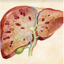
Liver
The liver is the largest internal organ and a metabolic powerhouse located in the upper right abdomen. It performs over 500 vital functions, including detoxification of metabolites, protein synthesis, production of biochemicals for digestion (bile), glycogen storage, regulation of blood glucose, synthesis of plasma proteins (e.g., albumin), and breakdown of old red blood cells. It has a remarkable capacity for regeneration.
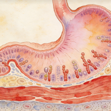
Stomach
The stomach is a J-shaped muscular sac in the upper abdomen that serves as a temporary reservoir for food. It mechanically churns and chemically digests food via gastric acid (HCl) and enzymes like pepsin. Its lining secretes mucus to protect itself. It regulates the delivery of partially digested food (chyme) into the small intestine. It also produces intrinsic factor, essential for vitamin B12 absorption.
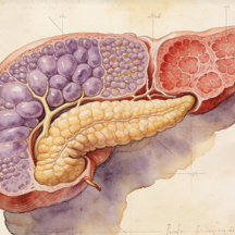
Pancreas
The pancreas is a dual-function gland located behind the stomach. Its exocrine portion (acinar cells) produces digestive enzymes (lipase, amylase, proteases) secreted into the duodenum. Its endocrine portion (islets of Langerhans) secretes hormones directly into the bloodstream, primarily insulin (from beta cells) and glucagon (from alpha cells), which are crucial for blood glucose regulation.
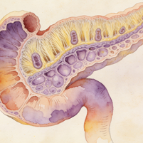
Intestine
The duodenum is the first and shortest segment (about 25 cm) of the small intestine, immediately following the stomach. It is a C-shaped structure where most chemical digestion occurs. It receives chyme from the stomach, bile from the liver/gallbladder, and digestive enzymes from the pancreas. Its Brunner's glands secrete alkaline mucus to neutralize gastric acid, protecting the intestinal lining and optimizing enzyme activity.
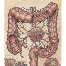
Intestine
The colon, or large intestine, is the final part of the digestive tract. Its main functions are to absorb water and electrolytes from indigestible food matter, compact the remaining material into feces, and store it until defecation. It houses a vast population of commensal bacteria (gut microbiota) that ferment certain substrates and produce vitamins (like K and some B vitamins). It consists of ascending, transverse, descending, and sigmoid sections.
Reproductive system
4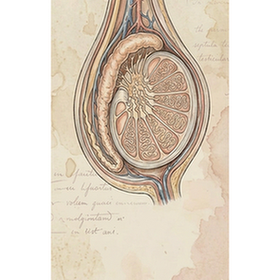
Testis
The testes (singular: testis) are the male primary gonads, housed in the scrotum. They have two primary functions: spermatogenesis, the production of spermatozoa in the seminiferous tubules; and steroidogenesis, the secretion of androgens, primarily testosterone, by Leydig cells. Temperature regulation in the scrotum is essential for normal spermatogenesis. Testosterone drives male secondary sexual characteristics and reproductive function.
Epididymis
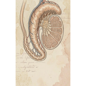
The epididymis is a tightly coiled, comma-shaped tube that sits on the posterior surface of each testis. It is the site of sperm maturation and storage. Immature sperm from the testes enter the head, gain motility and fertilizing capacity as they pass through the body, and are stored in the tail until ejaculation. Its secretions provide an environment for sperm to complete their functional development.
Ovary
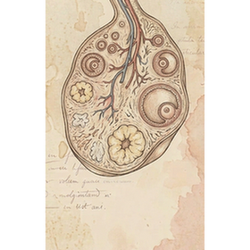
The ovaries are the female primary gonads, located in the pelvic cavity. They have two key functions: gametogenesis, the production and release of oocytes (eggs) through ovulation; and steroidogenesis, the secretion of the sex hormones estrogen and progesterone. These hormones regulate the menstrual cycle, support pregnancy, and maintain female secondary sexual characteristics. The ovaries contain a finite number of follicles at birth.
Uterus
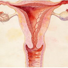
The uterus (womb) is a hollow, muscular, pear-shaped organ located in the female pelvis. It is the site of menstruation, implantation of a fertilized egg, gestation of the fetus, and labor. Its inner lining, the endometrium, cyclically thickens and sheds in response to ovarian hormones. Its thick myometrial wall contracts powerfully during childbirth. The cervix is its lower part, opening into the vagina.