Cell Types
Neural cells
13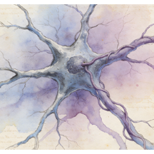
Astrocytes
Astrocytes are star-shaped glial cells, the most abundant in the central nervous system (CNS). They are vital for homeostasis: they form part of the blood-brain barrier, regulate blood flow, control extracellular ion/neurotransmitter balance (e.g., glutamate uptake), and provide metabolic support to neurons. Through the "tripartite synapse," they modulate synaptic strength and plasticity. They also respond to injury via reactive astrogliosis, playing dual roles in scar formation and neuroprotection.
Enteric glial cells
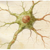
Enteric glial cells (EGCs) are the resident glia of the enteric nervous system (ENS), the "gut brain." They resemble CNS astrocytes and are integral to gastrointestinal homeostasis. EGCs support enteric neurons, regulate intestinal barrier integrity, modulate immune responses within the gut wall, and influence motility. Their dysfunction is implicated in inflammatory bowel diseases (IBD), infections, and functional GI disorders, highlighting their role beyond mere structural support.
Enteric neuronal progenitor cells
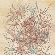
Enteric neuronal progenitor cells (ENPCs) are stem/precursor cells that give rise to the neurons and glia of the enteric nervous system (ENS). Primarily active during embryonic development, they migrate, proliferate, and differentiate to colonize the entire gut. Some progenitor-like cells may persist in adults, offering potential for ENS plasticity and repair. Defects in their migration cause Hirschsprung's disease, a congenital absence of ENS neurons in the distal colon.
Enteric neurons
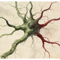
Enteric neurons are the functional units of the enteric nervous system (ENS), a complex network embedded in the gastrointestinal wall. They autonomously control gut functions including motility, secretion, blood flow, and immune responses. Comprising sensory neurons, interneurons, and motor neurons, they use diverse neurotransmitters (ACh, serotonin, NO, etc.). This "second brain" operates independently but communicates bidirectionally with the central nervous system.
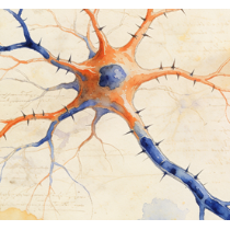
Excitatory neurons
Excitatory neurons are nerve cells that, when activated, increase the likelihood of their target neuron firing an action potential. They primarily release the neurotransmitter glutamate at their synapses. They form the primary driving force in neural circuits throughout the brain (e.g., cortical pyramidal neurons) and are fundamental for information propagation, synaptic plasticity (like LTP), and processes such as learning, memory, and cognition.
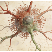
Granule cells
Granule cells are small, numerous excitatory neurons with a characteristic granular appearance. Key populations exist in the cerebellum (input neurons of the cortical layer), dentate gyrus of the hippocampus (forming new memories), and olfactory bulb. They typically receive multiple inputs, process information in a dense network, and project to larger principal cells (e.g., Purkinje cells, pyramidal cells), playing crucial roles in motor coordination, learning, and sensory processing.
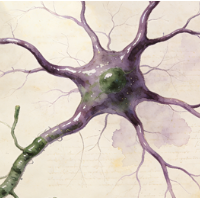
Inhibitory neurons
Inhibitory neurons reduce the probability of their postsynaptic target neuron firing an action potential. They primarily release the neurotransmitters GABA (in the brain) or glycine (in the spinal cord/brainstem). They are crucial for balancing neural circuit activity, preventing runaway excitation, generating rhythmic outputs (e.g., in pacemaker circuits), and shaping the temporal and spatial precision of neural computations. Types include interneurons like basket cells and Purkinje cells.
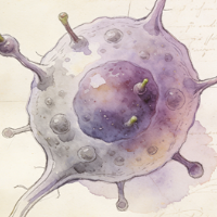
Oligodendrocyte progenitor cells
Oligodendrocyte progenitor cells (OPCs), also called NG2-glia, are a population of glial cells found throughout the CNS. They are proliferative and maintain the capacity to differentiate into mature, myelinating oligodendrocytes. OPCs are critical for developmental myelination and remain in the adult CNS, where they contribute to remyelination after demyelinating injuries (e.g., in Multiple Sclerosis) and may have additional roles in monitoring neural circuits.
Oligodendrocytes
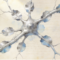
Oligodendrocytes are the myelinating glial cells of the central nervous system (CNS). Each oligodendrocyte extends multiple processes to wrap and insulate segments of several different axons with concentric layers of myelin membrane. This myelin sheath greatly increases the speed of action potential propagation (saltatory conduction). They also provide metabolic support to the axons they enwrap. Loss of oligodendrocytes or myelin disrupts nerve signaling.
Purkinje cells
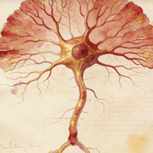
Purkinje cells are large, GABAergic inhibitory neurons that form a single layer in the cerebellar cortex. They are the sole output neurons of the cerebellar cortex, projecting to the deep cerebellar nuclei. They receive massive excitatory input from parallel fibers (granule cell axons) and powerful, instructive input from a single climbing fiber. Their complex dendritic tree integrates these signals to fine-tune motor coordination, balance, and motor learning.
Schwann cells
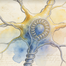
Schwann cells are the principal glial cells of the peripheral nervous system (PNS). Myelinating Schwann cells wrap around single axons to form the myelin sheath, enabling fast saltatory conduction. Non-myelinating Schwann cells ensheath multiple small-diameter axons. Schwann cells are crucial for supporting axon health and guiding regeneration after PNS injury, forming regeneration tubes (Bands of Büngner) that direct axonal regrowth.
Microglia
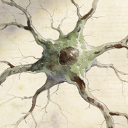
Microglia are the resident macrophages and primary immune cells of the central nervous system (CNS). In their resting state, they constantly survey the parenchyma. Upon detecting injury, infection, or pathological signals, they activate, becoming phagocytic to clear debris, dead cells, and pathogens. They also play essential roles in synaptic pruning during development, modulating neuroinflammation, and can contribute to both repair and neurodegeneration in diseases.
Chromaffin cells
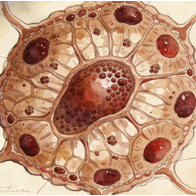
Chromaffin cells are neuroendocrine cells found primarily in the adrenal medulla. They are derived from neural crest cells and function as modified postganglionic sympathetic neurons. They synthesize and store catecholamines (epinephrine ~80%, norepinephrine ~20%) in dense-core secretory granules. In response to stress signals (acetylcholine from preganglionic fibers), they release these hormones directly into the bloodstream to mediate the systemic "fight-or-flight" response.
Immune cells
16
B cells
B cells are lymphocytes responsible for the humoral arm of the adaptive immune response. They develop in the bone marrow and, upon maturation, circulate in blood and lymph. When activated by antigen (often with T cell help), they proliferate and differentiate into antibody-secreting plasma cells or long-lived memory B cells. Their antibodies neutralize pathogens, opsonize them for phagocytosis, and activate the complement system.
Basophils
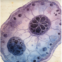
Basophils are the least abundant granulocyte in circulation. They contain large granules with histamine, heparin, and other mediators. They are primarily effector cells in allergic reactions and defense against parasites. When activated by IgE cross-linking or other stimuli, they degranulate, releasing inflammatory mediators that contribute to symptoms like vasodilation, itching, and bronchoconstriction. They also influence immune responses by secreting cytokines like IL-4.
Cycling B cells
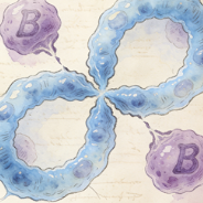
Cycling B cells refer to B lymphocytes that are actively proliferating, progressing through the cell cycle (G1, S, G2, M phases). This occurs during immune activation following antigen encounter and T cell help, leading to clonal expansion within germinal centers of lymph nodes or in extrafollicular sites. This proliferation is essential to generate a large population of antigen-specific B cell clones for antibody production and memory formation.
Cycling T cells
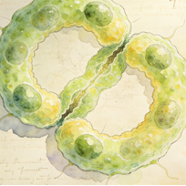
Cycling T cells are T lymphocytes undergoing active cell division. This is a hallmark of T cell activation following antigen recognition via the TCR and costimulation. Massive clonal expansion occurs, transforming a few naive antigen-specific T cells into thousands of effector T cells to combat infection. It is a fundamental step in mounting an effective adaptive immune response and generating long-lived memory T cells.
Dendritic cells
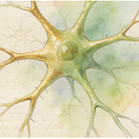
Dendritic cells (DCs) are professional antigen-presenting cells (APCs) that act as sentinels linking innate and adaptive immunity. Immature DCs in tissues capture antigens, then mature and migrate to lymph nodes. There, they present processed antigens on MHC molecules to naive T cells, providing the critical "signal 1" and costimulatory "signal 2" required for T cell activation and differentiation, thereby initiating antigen-specific immune responses.
Kupffer cells
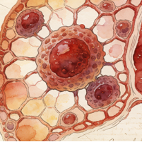
Kupffer cells are specialized tissue-resident macrophages located within the sinusoids of the liver. They are the largest population of fixed macrophages in the body. They phagocytose pathogens, toxins, cellular debris, and aged red blood cells from the portal blood flow. They play crucial roles in liver immunity, iron recycling, lipid metabolism, and maintaining overall hepatic homeostasis. Their activation can contribute to liver inflammation and fibrosis.
Macrophages
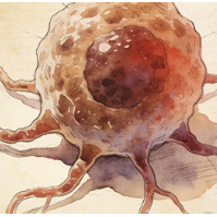
Macrophages are large, phagocytic cells of the innate immune system present in all tissues (where they have specific names like Kupffer cells, microglia). They derive from blood monocytes or local progenitors. They engulf and destroy pathogens and dead cells, secrete cytokines and chemokines to regulate inflammation, present antigens, and are key players in tissue repair, remodeling, and homeostasis. They exhibit remarkable functional plasticity (M1/M2 spectra).
Mast cells
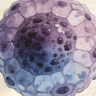
Mast cells are tissue-resident granulocytes found near blood vessels and nerves, particularly in skin, lungs, and gut mucosa. They store pre-formed inflammatory mediators (histamine, tryptase, heparin) in their granules. They are central effectors in IgE-mediated allergic reactions (anaphylaxis, hay fever) and defense against parasites. Upon activation, they rapidly degranulate and also synthesize cytokines/chemokines, influencing inflammation, immunity, and even tissue remodeling.
Monocytes
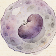
Monocytes are circulating white blood cells (agranulocytes) that serve as precursors for macrophages and dendritic cells. Produced in the bone marrow, they patrol the bloodstream for several days before migrating into tissues in response to inflammatory signals. In tissues, they differentiate into macrophages or dendritic cells. In blood, they can phagocytose pathogens and present antigens, acting as a bridge between innate and adaptive immunity.
Natural killer cells
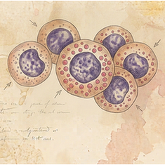
Natural Killer (NK) cells are cytotoxic lymphocytes of the innate immune system. They provide rapid responses to virus-infected cells and tumor cells without prior sensitization. They use a balance of activating and inhibitory receptors to detect "missing self" (loss of MHC I) or "induced self" (stress ligands). They kill targets by releasing perforin and granzymes (cytotoxic granules) and through death receptor ligands like FasL.
Natural killer T cells
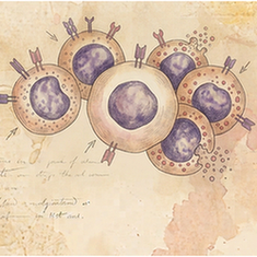
Natural Killer T (NKT) cells are a unique subset of T lymphocytes that bridge innate and adaptive immunity. They express an invariant T cell receptor (TCR) that recognizes lipid antigens presented by the non-classical MHC molecule CD1d. Upon activation, they rapidly produce large quantities of cytokines (e.g., IFN-γ, IL-4), influencing the activity of many other immune cells and shaping immune responses to infection, cancer, and autoimmunity.
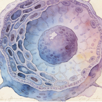
Plasma B cells
Plasma B cells (or plasma cells) are the terminal, fully differentiated effector state of activated B lymphocytes. They are antibody factories, specializing in the massive secretion of antibodies of a single specificity. They possess extensive endoplasmic reticulum and a prominent Golgi apparatus to support high-rate protein synthesis. Most are short-lived, but some become long-lived plasma cells that reside in the bone marrow, providing sustained antibody levels.
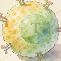
T cells
T cells (T lymphocytes) are central players in cell-mediated adaptive immunity. They develop in the thymus and express T cell receptors (TCRs) that recognize peptide antigens presented by MHC molecules. Major subsets include: Helper T cells (CD4+, which secrete cytokines to help B cells and macrophages), Cytotoxic T cells (CD8+, which kill infected/cancerous cells), and Regulatory T cells (Tregs, which suppress immune responses to prevent autoimmunity).
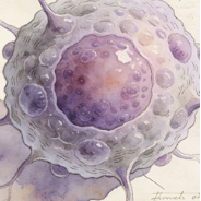
Thymocytes
Thymocytes are developing T cell precursors within the thymus. They originate from bone marrow-derived progenitors that migrate to the thymus. Here, they undergo a rigorous process of maturation involving proliferation, TCR gene rearrangement, and positive/negative selection. Only those with a functional TCR that recognizes self-MHC with moderate affinity (positive selection) but not self-antigens too strongly (negative selection) survive to become mature, naive T cells.
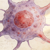
Goblet cells
Goblet cells are specialized unicellular mucus-secreting glands found within simple columnar epithelia of the respiratory and intestinal tracts. They have a characteristic goblet shape, with a basal nucleus and an apical region packed with mucin granules. They secrete mucins, which hydrate and form the protective mucus layer that traps particles, lubricates surfaces, and protects the epithelium from pathogens and damage.
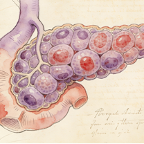
Pancreas islet cells
Pancreatic islet (islets of Langerhans) cells are the endocrine cells of the pancreas, organized into clusters scattered throughout the exocrine tissue. Major types include: Alpha cells (secrete glucagon, raises blood sugar), Beta cells (secrete insulin, lowers blood sugar), Delta cells (secrete somatostatin, inhibits other islet cells), and PP cells (secrete pancreatic polypeptide). They precisely regulate glucose homeostasis.
Epithelial cells
32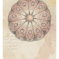
Acinar cells
Acinar cells are the exocrine secretory cells of glands like the salivary glands and pancreas. In the pancreas, they are organized into acini and synthesize, store, and secrete digestive pro-enzymes (zymogens) into a ductal network. They have a highly developed rough ER and abundant secretory (zymogen) granules. When stimulated (e.g., by cholecystokinin), they release their contents into the pancreatic duct, which empties into the duodenum.
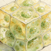
Alveolar type 2 cells
Alveolar type 2 (AT2) cells are cuboidal epithelial cells found in the alveolar corners. They have three critical functions: (1) They synthesize and secrete pulmonary surfactant, a phospholipid-protein mixture that reduces surface tension and prevents alveolar collapse. (2) They serve as progenitor cells for both AT1 and AT2 cells, repairing the alveolar epithelium after injury. (3) They contribute to innate immune defense in the alveoli.
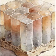
Basal cells
Basal cells are a layer of mitotically active epithelial progenitor/stem cells found attached to the basement membrane in stratified and pseudostratified epithelia (e.g., skin, airways, prostate). They divide to give rise to cells that differentiate and move upward to replenish the superficial layers. In the airway, they are major stem cells capable of regenerating both ciliated and secretory cells after injury.
Cholangiocytes
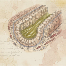
Cholangiocytes are the epithelial cells that line the intrahepatic and extrahepatic bile ducts. They modify the composition of bile (through secretion and absorption of water, ions, and bicarbonate), form a protective barrier, and respond to hormonal signals. They express receptors and channels (e.g., CFTR) critical for bile flow. Their proliferation and dysfunction are central to cholangiopathies like primary sclerosing cholangitis (PSC).
Ciliated cells
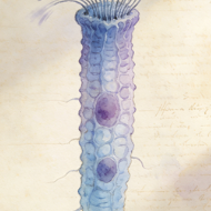
Ciliated cells are epithelial cells characterized by the presence of motile cilia (hair-like projections) on their apical surface. They are found in the respiratory tract, oviducts, and ventricles of the brain (ependymal cells). Coordinated, rhythmic beating of cilia moves fluid or mucus over the epithelial surface. In the airways, this "mucociliary escalator" traps and propels inhaled particles and pathogens out of the lungs, a key defense mechanism.
Cycling epithelial cells
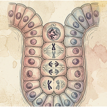
A broad categorization for any epithelial cell (from various tissues) that is captured in an active phase of the cell cycle. This label is used in genomic datasets to distinguish proliferating cells from quiescent or terminally differentiated ones. It highlights the dynamic nature of epithelia, where constant renewal and repair are driven by these cycling populations.
Cycling gastric epithelial cell
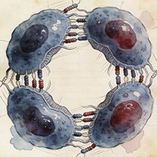
Cycling gastric epithelial cell (Epithelial cells) in species SP013. Single-cell UMAP atlas.
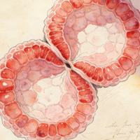
Cycling luminal epithelial cells
Luminal epithelial cells line the inner (luminal) surface of ducts and glands (e.g., in mammary gland, prostate). "Cycling" identifies those that are in the cell cycle. In hormone-responsive tissues, the proportion of cycling luminal cells can fluctuate with hormonal signals. They may represent a progenitor population or mature cells undergoing renewal.
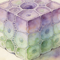
Distal convoluted tubule cells
Cells of the distal convoluted tubule (DCT) in the nephron. They are responsible for fine-tuning electrolyte balance. They actively reabsorb sodium and chloride (via the Na-Cl cotransporter, NCC) and are a major site for parathyroid hormone (PTH)-regulated calcium reabsorption. Their activity is crucial for maintaining blood pressure, potassium, calcium, and magnesium homeostasis.
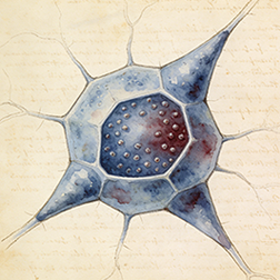
Endocrine cells
Endocrine cells (Epithelial cells) in species SP013. Single-cell UMAP atlas.
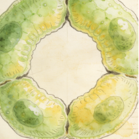
Enterocytes
Enterocytes are the principal absorptive columnar epithelial cells lining the small intestine villi. Their apical surface is covered with microvilli, forming a "brush border" that dramatically increases surface area for nutrient absorption. They digest and absorb carbohydrates, peptides, amino acids, and lipids. They also secrete water and ions and form a crucial barrier between the gut lumen and the internal environment.
Enteroendocrine cells
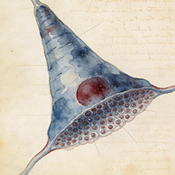
Enteroendocrine cells (Epithelial cells) in species SP013. Single-cell UMAP atlas.
Fasciculata cells
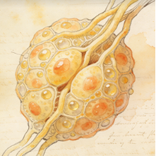
Fasciculata cells are steroidogenic endocrine cells that constitute the zona fasciculata, the middle and largest layer of the adrenal cortex. Their primary function is the production of glucocorticoids (mainly cortisol in humans), stimulated by ACTH from the pituitary. They contain abundant lipid droplets with cholesterol esters (giving a "foamy" appearance) and smooth endoplasmic reticulum for steroid hormone synthesis.
Follicular cells
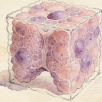
In the thyroid gland, follicular cells (thyrocytes) are the principal epithelial cells that form spherical follicles. They synthesize thyroglobulin and, upon stimulation by TSH, take up iodine to produce the thyroid hormones thyroxine (T4) and triiodothyronine (T3), which are stored in the colloid-filled follicle lumen and released into the blood to regulate metabolism, growth, and development.
Glandular cells
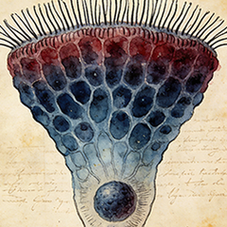
Glandular cells are specialized epithelial cells that constitute the secretory units of exocrine and endocrine glands. They synthesize and release specific products. Exocrine glandular cells (e.g., in salivary, sweat glands) secrete via ducts. Endocrine glandular cells (e.g., in thyroid, pituitary) secrete hormones directly into the bloodstream. Their morphology is adapted for secretion, with abundant rough ER and secretory granules.
Glomerulosa cells
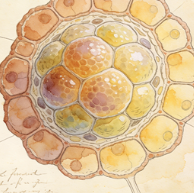
Glomerulosa cells are steroid-producing endocrine cells that form the outermost zona glomerulosa of the adrenal cortex. They are uniquely capable of producing the mineralocorticoid aldosterone, regulated primarily by the renin-angiotensin-aldosterone system (RAAS) and potassium levels. Aldosterone acts on the kidneys to promote sodium retention and potassium excretion, regulating blood pressure and electrolyte balance.
Gonadotropes
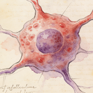
Gonadotropes are endocrine cells in the anterior pituitary gland that produce and secrete the gonadotropins: follicle-stimulating hormone (FSH) and luteinizing hormone (LH). In response to gonadotropin-releasing hormone (GnRH) from the hypothalamus, they release these hormones into the bloodstream. FSH and LH then act on the gonads (ovaries and testes) to regulate gametogenesis and sex steroid hormone production.
Hepatocytes
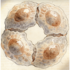
Hepatocytes are the principal parenchymal cells of the liver, making up about 80% of its mass. They are metabolic powerhouses with functions including: protein synthesis (albumin, clotting factors), bile production, detoxification and metabolism of drugs/toxins, glycogen storage and glucose homeostasis, lipid metabolism, and cholesterol synthesis. They are arranged in plates radiating from central veins, with polarized faces facing sinusoids and bile canaliculi.
Keratinocytes
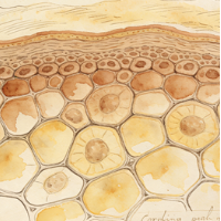
Keratinocytes are the predominant cell type in the epidermis (the outermost skin layer). They originate from basal layer stem cells and undergo a process of terminal differentiation as they move outward, producing keratin intermediate filaments. In the stratum corneum, they become anucleate corneocytes, forming a tough, waterproof barrier that protects against mechanical injury, water loss, and microbial invasion.
Lactotropes
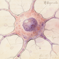
Lactotropes (or mammotropes) are endocrine cells in the anterior pituitary gland that produce and secrete prolactin (PRL). Prolactin's primary role is to stimulate milk production (lactation) in the mammary glands after childbirth. Its secretion is primarily under inhibitory control by dopamine from the hypothalamus. Lactotropes can increase in number during pregnancy and lactation.
Luminal epithelial cells
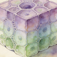
These are the epithelial cells that directly face the lumen (hollow cavity) of a duct or gland. In tissues like the mammary gland or prostate, they form the inner secretory layer and are often hormonally responsive. They are typically more differentiated than basal cells and carry out the tissue-specific secretory functions (e.g., producing milk proteins or prostatic fluid).
Mucous cells
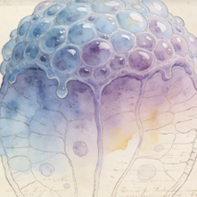
Mucous cells are specialized secretory epithelial cells that produce and secrete mucus (mucins). They are found in the surface epithelium of the stomach, in salivary glands (as mucous acinar cells), and elsewhere. Their secretions serve to lubricate, protect, and hydrate epithelial surfaces. The mucin granules often stain lightly with H&E, giving the cytoplasm a clear or foamy appearance.
Neuroendocrine cells
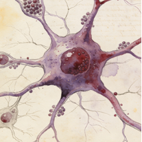
Neuroendocrine cells are specialized cells that possess properties of both neurons (they receive neural input and may contain dense-core vesicles) and endocrine cells (they secrete hormones or peptides into the bloodstream). They are found scattered in various epithelia (e.g., lungs, GI tract) and in dedicated glands (adrenal medulla, pituitary). They release substances like serotonin, gastrin, or catecholamines in response to neuronal or chemical stimuli.
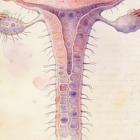
Oviduct epithelial cells
These cells line the fallopian tubes (oviducts). The epithelium is simple columnar, consisting of two main cell types: ciliated cells, whose beating helps move the egg/embryo toward the uterus, and secretory (peg) cells, which secrete substances that nourish gametes and facilitate fertilization and early embryo development. Their function is hormonally regulated and critical for reproduction.
Pancreas ductal cells
Pancreatic ductal cells form the lining of the ductal network that transports digestive enzymes from the acini and bicarbonate-rich fluid from the centroacinar cells to the duodenum. They modify the pancreatic juice by secreting bicarbonate ions (HCO3-) to neutralize gastric acid. They express CFTR, and mutations here cause cystic fibrosis-related pancreatic disease. They can also act as progenitor cells in certain contexts.
Parietal cells
Parietal cells (or oxyntic cells) are found in the gastric glands of the stomach lining. They secrete hydrochloric acid (HCl) into the stomach lumen, creating the highly acidic environment necessary for protein digestion and killing ingested microbes. They also secrete intrinsic factor, a glycoprotein essential for vitamin B12 absorption in the ileum. They have a distinctive intracellular canaliculus system for acid secretion.
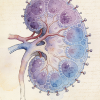
Podocytes
Podocytes are highly specialized epithelial cells in the Bowman's capsule of the kidney glomerulus. They wrap around capillaries with primary and secondary foot processes. The slits between these foot processes, covered by the slit diaphragm, form the final filtration barrier preventing proteins from entering the urine. Podocyte injury or loss leads to proteinuria and glomerular diseases like focal segmental glomerulosclerosis (FSGS).
Principal cells
In the kidney's collecting duct, principal cells are the most abundant cell type. They are responsible for the final regulation of water, sodium, and potassium balance. They express aquaporin-2 channels on their apical membrane (inserted in response to vasopressin/ADH) for water reabsorption. They also reabsorb sodium via ENaC channels and secrete potassium, processes regulated by aldosterone.
Progenitor-like epithelial cells
This describes epithelial cells that exhibit molecular signatures of progenitor or stem cells but are not the most primitive stem cell. They are often in an intermediate state, possessing the capacity to proliferate and give rise to differentiated cells, but may have some lineage restriction. They are crucial for steady-state tissue maintenance and repair after injury.
Proximal tubule cells
These cells line the proximal convoluted tubule (PCT) of the nephron, the first and longest segment. They are responsible for the bulk reabsorption of filtered nutrients (glucose, amino acids), ions (sodium, chloride, bicarbonate), and water (~65%). Their apical surface has a dense brush border of microvilli to increase surface area. They also actively secrete organic acids and bases into the filtrate.
Serous cells
Serous cells are protein-secreting epithelial cells found in exocrine glands like the salivary glands (parotid) and pancreatic acini. Their secretory granules contain enzymes (e.g., amylase in saliva, digestive enzymes in pancreas). The granules often stain darkly with H&E. They typically have a round nucleus and basophilic cytoplasm due to abundant rough ER for protein synthesis.
Taste cells
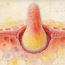
Taste cells are specialized sensory epithelial cells grouped into taste buds on the tongue and palate. They are not neurons but are chemosensory receptors that synapse with afferent nerve fibers. Different types detect sweet, salty, sour, bitter, and umami (savory) stimuli. When activated, they depolarize and release neurotransmitters to stimulate gustatory nerves, sending signals to the brain.
Stromal cells
8
Adipocyte progenitor cells
Adipocyte progenitor cells (APCs), also known as preadipocytes, are mesenchymal stem cells committed to the adipocyte lineage. They reside in adipose tissue depots and have the capacity to proliferate and differentiate into mature lipid-filled adipocytes. They play key roles in adipose tissue expansion, both in normal growth and in obesity. They also secrete various factors (adipokines) and contribute to tissue remodeling.
Fibroblasts
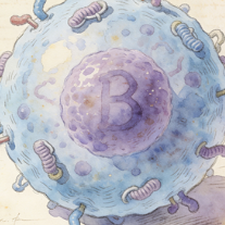
Fibroblasts are the most common cells of connective tissue. They synthesize and secrete the extracellular matrix (ECM) components, including collagen, elastin, and fibronectin, providing structural and biochemical support to surrounding cells. They are key players in wound healing, where they proliferate, migrate to the injury site, and differentiate into contractile myofibroblasts to close wounds. They also communicate with immune and epithelial cells.
Folliculostellate cells
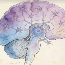
Folliculostellate cells are non-endocrine, sustentacular-like cells found in the anterior pituitary gland. They form a network surrounding the hormone-producing endocrine cells. They produce growth factors and cytokines, regulate blood flow and the local microenvironment, phagocytose debris, and may coordinate the activity of pituitary endocrine cells through gap junctions and paracrine signaling.
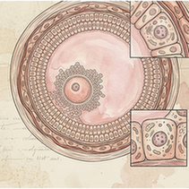
Granulosa cells
Granulosa cells are the somatic cells that surround and support the developing oocyte within the ovarian follicle. They produce estrogen (from androgen precursors supplied by theca cells) and inhibin. They also supply nutrients to the oocyte and communicate with it via gap junctions. After ovulation, they transform into luteal cells of the corpus luteum, which secrete progesterone.
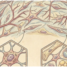
Mesenchymal cells
Mesenchymal cells are a broad category of cells derived from the mesoderm. They include fibroblasts, adipocytes, osteoblasts, chondrocytes, and muscle cells. In development, they give rise to connective tissues, bone, cartilage, and blood vessels. Mesenchymal stem/stromal cells (MSCs) are multipotent adult cells that can differentiate into these lineages and have immunomodulatory properties, holding potential for regenerative medicine.
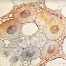
Other stromal cells
Stromal cells are a heterogeneous group of connective tissue cells that provide structural support and create the microenvironment ("stroma") for parenchymal cells within an organ. They include fibroblasts, adipocytes, pericytes, and various types of mesenchymal stem/stromal cells. They produce ECM, secrete growth factors and cytokines, and play critical roles in tissue architecture, homeostasis, repair, and disease progression (e.g., in cancer).
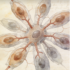
Pericytes
Pericytes are contractile mesenchymal cells that wrap around the endothelial cells of capillaries and venules. They are embedded in the basement membrane and are crucial for vascular stability, regulating blood flow, promoting endothelial survival, and participating in the formation and maintenance of the blood-brain barrier. They also have stem cell properties and can differentiate into other mesenchymal cells during tissue repair.
Stellate cells
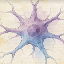
In the liver, hepatic stellate cells (HSCs) reside in the space of Disse. In their quiescent state, they store vitamin A. Upon liver injury, they activate, proliferate, and transform into myofibroblast-like cells that produce large amounts of extracellular matrix, driving liver fibrosis and cirrhosis. Pancreatic stellate cells play a similar fibrogenic role in pancreatitis and pancreatic cancer.
Muscle cells
2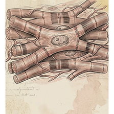
Cardiomyocytes
Cardiomyocytes are the striated muscle cells that constitute the cardiac muscle of the heart. They are responsible for generating the contractile force that pumps blood. They are connected end-to-end by intercalated discs containing gap junctions for electrical coupling and desmosomes for mechanical attachment. Unlike skeletal muscle, they are mononucleated or binucleated and have intrinsic automaticity (pacemaker cells).
Smooth muscle cells
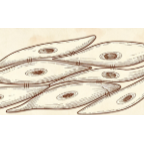
Smooth muscle cells (SMCs) are involuntary, non-striated muscle cells found in the walls of hollow organs (e.g., blood vessels, gut, bladder, uterus). They are spindle-shaped with a single central nucleus. They contract slowly and can maintain tension for long periods (tonus). Contraction is regulated by the autonomic nervous system, hormones, and local factors (e.g., nitric oxide, endothelin).
Other
3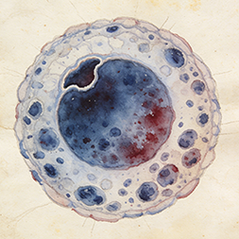
Myelocytes
Myelocytes (Other) in species SP013. Single-cell UMAP atlas.
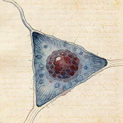
Colonic stem cells
Colonic stem cells (Other) in species SP013. Single-cell UMAP atlas.
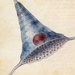
Intercalated cells
Intercalated cells are specialized cells found in the collecting ducts of the kidney nephron. They are critical for regulating systemic acid-base balance. There are two main types: Type A (alpha) cells secrete protons (H+) via H+-ATPase pumps and reabsorb bicarbonate, correcting acidosis. Type B (beta) cells secrete bicarbonate and reabsorb protons, correcting alkalosis. They also play a role in potassium balance.