Cell Types
Neural cells
2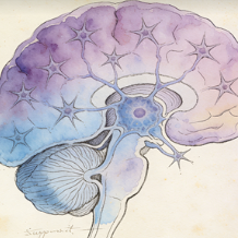
Immune cells
7
B cells
B cells are lymphocytes responsible for the humoral arm of the adaptive immune response. They develop in the bone marrow and, upon maturation, circulate in blood and lymph. When activated by antigen (often with T cell help), they proliferate and differentiate into antibody-secreting plasma cells or long-lived memory B cells. Their antibodies neutralize pathogens, opsonize them for phagocytosis, and activate the complement system.
Dendritic cells
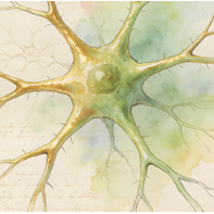
Dendritic cells (DCs) are professional antigen-presenting cells (APCs) that act as sentinels linking innate and adaptive immunity. Immature DCs in tissues capture antigens, then mature and migrate to lymph nodes. There, they present processed antigens on MHC molecules to naive T cells, providing the critical "signal 1" and costimulatory "signal 2" required for T cell activation and differentiation, thereby initiating antigen-specific immune responses.
Kupffer cells
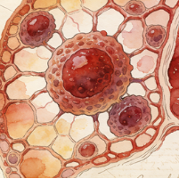
Kupffer cells are specialized tissue-resident macrophages located within the sinusoids of the liver. They are the largest population of fixed macrophages in the body. They phagocytose pathogens, toxins, cellular debris, and aged red blood cells from the portal blood flow. They play crucial roles in liver immunity, iron recycling, lipid metabolism, and maintaining overall hepatic homeostasis. Their activation can contribute to liver inflammation and fibrosis.
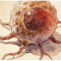
Macrophages
Macrophages are large, phagocytic cells of the innate immune system present in all tissues (where they have specific names like Kupffer cells, microglia). They derive from blood monocytes or local progenitors. They engulf and destroy pathogens and dead cells, secrete cytokines and chemokines to regulate inflammation, present antigens, and are key players in tissue repair, remodeling, and homeostasis. They exhibit remarkable functional plasticity (M1/M2 spectra).
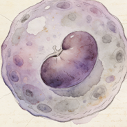
Monocytes
Monocytes are circulating white blood cells (agranulocytes) that serve as precursors for macrophages and dendritic cells. Produced in the bone marrow, they patrol the bloodstream for several days before migrating into tissues in response to inflammatory signals. In tissues, they differentiate into macrophages or dendritic cells. In blood, they can phagocytose pathogens and present antigens, acting as a bridge between innate and adaptive immunity.
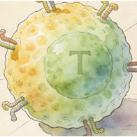
T cells
T cells (T lymphocytes) are central players in cell-mediated adaptive immunity. They develop in the thymus and express T cell receptors (TCRs) that recognize peptide antigens presented by MHC molecules. Major subsets include: Helper T cells (CD4+, which secrete cytokines to help B cells and macrophages), Cytotoxic T cells (CD8+, which kill infected/cancerous cells), and Regulatory T cells (Tregs, which suppress immune responses to prevent autoimmunity).
Immune cells
Immune cells (Immune cells) in species SP113. Single-cell UMAP atlas.
Epithelial cells
9
Alveolar type 1 cells
Alveolar type 1 (AT1) cells are thin, squamous epithelial cells that cover approximately 95% of the gas exchange surface in the lung alveoli. Their extremely flattened morphology (extending over large areas) is ideal for the passive diffusion of oxygen and carbon dioxide between the airspace and the underlying pulmonary capillaries. They are terminally differentiated and crucial for maintaining the delicate blood-air barrier.
Alveolar type 2 cells
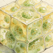
Alveolar type 2 (AT2) cells are cuboidal epithelial cells found in the alveolar corners. They have three critical functions: (1) They synthesize and secrete pulmonary surfactant, a phospholipid-protein mixture that reduces surface tension and prevents alveolar collapse. (2) They serve as progenitor cells for both AT1 and AT2 cells, repairing the alveolar epithelium after injury. (3) They contribute to innate immune defense in the alveoli.
Ascending loop of Henle cells
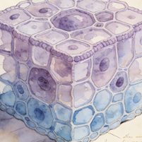
Cells of the thick ascending limb (TAL) of the loop of Henle in the kidney nephron. They are impermeable to water but actively reabsorb sodium, potassium, and chloride via the Na-K-2Cl cotransporter (NKCC2) on their apical membrane. This transport is critical for generating the hypertonic medullary interstitium necessary for water reabsorption in the collecting duct. They also contribute to magnesium and calcium reabsorption.
Cholangiocytes
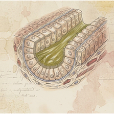
Cholangiocytes are the epithelial cells that line the intrahepatic and extrahepatic bile ducts. They modify the composition of bile (through secretion and absorption of water, ions, and bicarbonate), form a protective barrier, and respond to hormonal signals. They express receptors and channels (e.g., CFTR) critical for bile flow. Their proliferation and dysfunction are central to cholangiopathies like primary sclerosing cholangitis (PSC).
Ciliated cells
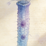
Ciliated cells are epithelial cells characterized by the presence of motile cilia (hair-like projections) on their apical surface. They are found in the respiratory tract, oviducts, and ventricles of the brain (ependymal cells). Coordinated, rhythmic beating of cilia moves fluid or mucus over the epithelial surface. In the airways, this "mucociliary escalator" traps and propels inhaled particles and pathogens out of the lungs, a key defense mechanism.
Connecting tubule cells
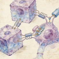
Connecting tubule (CNT) cells are specialized cells in the nephron that link the distal convoluted tubule to the collecting duct. They play a significant role in fine-tuning sodium, potassium, and acid-base balance. They are sensitive to aldosterone and reabsorb sodium while secreting potassium and protons. They express the calcium channel TRPV5 and are important for the final regulation of calcium reabsorption.
Hepatocytes
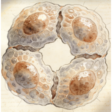
Hepatocytes are the principal parenchymal cells of the liver, making up about 80% of its mass. They are metabolic powerhouses with functions including: protein synthesis (albumin, clotting factors), bile production, detoxification and metabolism of drugs/toxins, glycogen storage and glucose homeostasis, lipid metabolism, and cholesterol synthesis. They are arranged in plates radiating from central veins, with polarized faces facing sinusoids and bile canaliculi.
Principal cells
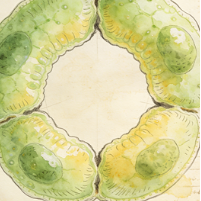
In the kidney's collecting duct, principal cells are the most abundant cell type. They are responsible for the final regulation of water, sodium, and potassium balance. They express aquaporin-2 channels on their apical membrane (inserted in response to vasopressin/ADH) for water reabsorption. They also reabsorb sodium via ENaC channels and secrete potassium, processes regulated by aldosterone.
Proximal tubule cells
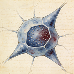
These cells line the proximal convoluted tubule (PCT) of the nephron, the first and longest segment. They are responsible for the bulk reabsorption of filtered nutrients (glucose, amino acids), ions (sodium, chloride, bicarbonate), and water (~65%). Their apical surface has a dense brush border of microvilli to increase surface area. They also actively secrete organic acids and bases into the filtrate.
Stromal cells
3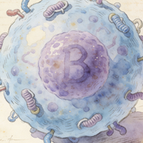
Fibroblasts
Fibroblasts are the most common cells of connective tissue. They synthesize and secrete the extracellular matrix (ECM) components, including collagen, elastin, and fibronectin, providing structural and biochemical support to surrounding cells. They are key players in wound healing, where they proliferate, migrate to the injury site, and differentiate into contractile myofibroblasts to close wounds. They also communicate with immune and epithelial cells.
Stellate cells
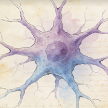
In the liver, hepatic stellate cells (HSCs) reside in the space of Disse. In their quiescent state, they store vitamin A. Upon liver injury, they activate, proliferate, and transform into myofibroblast-like cells that produce large amounts of extracellular matrix, driving liver fibrosis and cirrhosis. Pancreatic stellate cells play a similar fibrogenic role in pancreatitis and pancreatic cancer.
Stromal cells
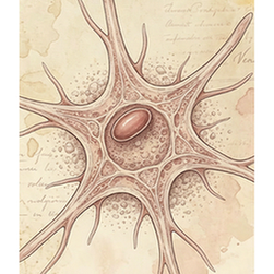
Stromal cells in human. 101,901 cells across 16 tissues.
Muscle cells
2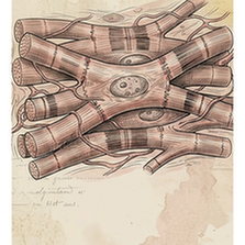
Cardiomyocytes
Cardiomyocytes are the striated muscle cells that constitute the cardiac muscle of the heart. They are responsible for generating the contractile force that pumps blood. They are connected end-to-end by intercalated discs containing gap junctions for electrical coupling and desmosomes for mechanical attachment. Unlike skeletal muscle, they are mononucleated or binucleated and have intrinsic automaticity (pacemaker cells).
Smooth muscle cells
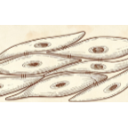
Smooth muscle cells (SMCs) are involuntary, non-striated muscle cells found in the walls of hollow organs (e.g., blood vessels, gut, bladder, uterus). They are spindle-shaped with a single central nucleus. They contract slowly and can maintain tension for long periods (tonus). Contraction is regulated by the autonomic nervous system, hormones, and local factors (e.g., nitric oxide, endothelin).
Other
5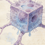
Mesothelial cells
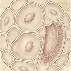
Mesothelial cells form a monolayer (the mesothelium) that lines the body's serous cavities (pleural, pericardial, peritoneal) and covers the outer surface of internal organs. They produce a lubricating serous fluid that allows organs to move smoothly. They also provide a protective barrier, participate in immune surveillance and inflammation, and can undergo transition to a fibroblast-like phenotype (mesothelial-to-mesenchymal transition) in pathology.
Proliferating cells
Proliferating cells (Other) in species SP113. Single-cell UMAP atlas.
Unknown cell type
Unknown cell type (Other) in species SP113. Single-cell UMAP atlas.
Pluripotent stem cells
Pluripotent stem cells (Other) in species SP113. Single-cell UMAP atlas.
Cycling cells
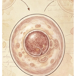
Cycling cells are cells actively progressing through the cell cycle (G1, S, G2, and M phases), typically identified by expression of proliferation markers such as MKI67 and TOP2A. In single-cell atlases they represent a transient proliferative state rather than a fixed lineage and can arise from multiple progenitor or stem cell populations within a tissue. Their abundance reflects the turnover and regenerative activity of the tissue sampled.